40 meniscus diagram of the knee
Gross anatomy — Gross anatomy. There are two fibrocartilaginous menisci in the knee joint: a medial meniscus within the medial tibiofemoral compartment and ...Red-white zone: transition between outer third ...Red zone: outer one-thirdWhite zone: inner two-thirds Anatomy of Menisci · by AJS Fox · 2012 · Cited by 355 — Gross Anatomy. Gross examination of the knee menisci reveals a smooth, lubricated tissue (Figure 1).
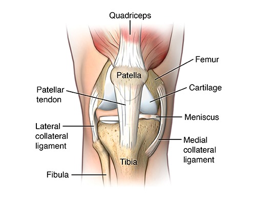
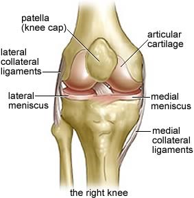
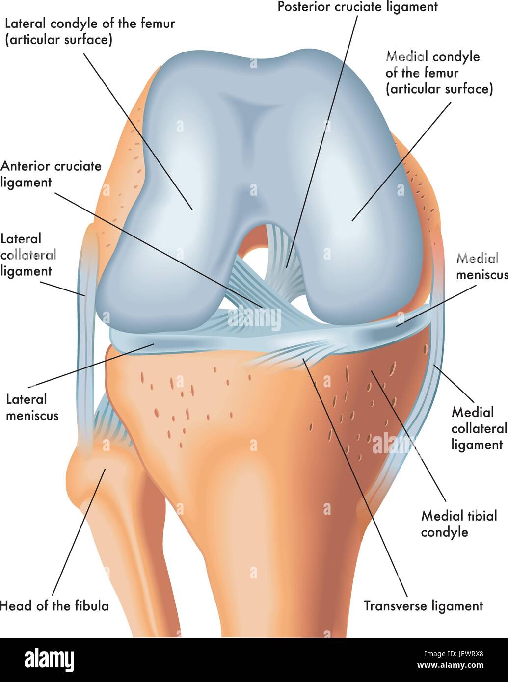
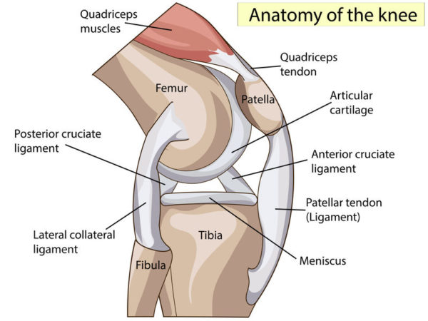
Articular cartilage lines the joint surfaces of the bones in the knee (tibia, femur, and patella, or kneecap). The medial and lateral meniscus are two thicker ...

Meniscus diagram of the knee
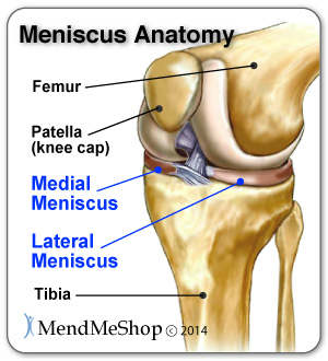
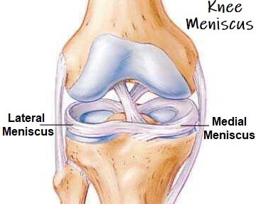
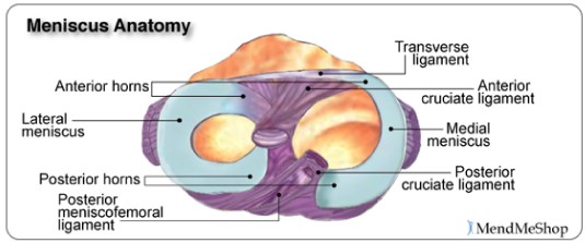
Diagram of Knee Joint showing Meniscus The meniscus is a crescent-shaped structure composed of cartilage that functions to distribute body weight evenly across the three bones that make up the knee joint: the thigh bone, shin bone, and knee cap. Knee pain could be the result of a problem with any one of these components, or a combination of several. You may be experiencing knee pain and want to know the possible causes. The diagram, below, is a handy guide to the possible reasons for your pain. Pain at the front above the knee We are pleased to provide you with the picture named Meniscus anatomy diagram.We hope this picture Meniscus anatomy diagram can help you study and research. for more anatomy content please follow us and visit our website: www.anatomynote.com. Anatomynote.com found Meniscus anatomy diagram from plenty of anatomical pictures on the internet.We think this is the most useful anatomy picture that ...
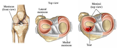
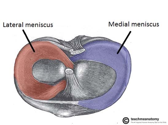
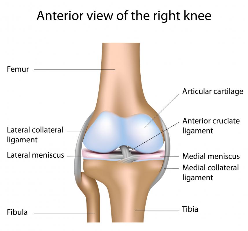
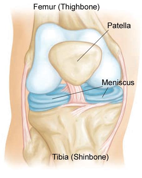
Meniscus diagram of the knee. Medial meniscus. The medial meniscus is the central band of cartilage attached to the tibia, or shinbone. The band goes around the knee joint in a crescent-shaped path and is located between the ... The knee is the joint where the large bones of the upper leg (femur), lower leg (tibia), and kneecap ( patella) meet. Two pieces of “C-shaped” cartilage called the meniscus cushion the ends of the femur and tibia at the knee joint. A tear in this cartilage is called a meniscal tear. Meniscal tears have different names – longitudinal, flap ... 2.1 Meniscus Anatomy. The knee joint contains the meniscus structure, comprised of both a medial and a lateral component situated between the corresponding femoral condyle and tibial plateau (Figure 1) [].Each is a glossy-white, complex tissue comprised of cells, specialized extracellular matrix (ECM) molecules, and region-specific innervation and vascularization. The meniscus is a rubber-like C-shaped disc that sits within the knee between the end of the thighbone (femur) and the top of the shinbone (tibia). In the ...
This first knee pain diagnosis chart focuses on pain at the front of the knee. Then next one, further down, looks at pain behind the knee. A. Pain Above the Knee Cap (yellow). Quadriceps Tendinopathy: Damage to the quadriceps tendon causing pain above the kneecap that is worse with activity. LEARN MORE> B. Outer Knee Pain (blue). Iliotibial Band Syndrome: Most common. Menisci rests between the thigh bone femur and the tibia and there are two knee joint ligaments. They are a type of cartilage in the joint. The rubbery texture ...Introduction · Anatomy and attachment · Injury/Tear · Diagnosis The knee is a complex joint that flexes, extends, and twists slightly from side to side. The knee is the meeting point of the femur (thigh bone) in the upper leg and the tibia (shinbone) in the ... 15+ Meniscus Diagram Of The Knee Pictures. Indicate the amount of meniscus that was excised by drawing on the diagram and. Free access interactive and dynamic anatomical atlas. Knee Meniscus Anatomy from mendmyknee.com. It consists of bones, meniscus, ligaments, and tendons. It may also be referred to as a bicondylar joint as movement primarily ...
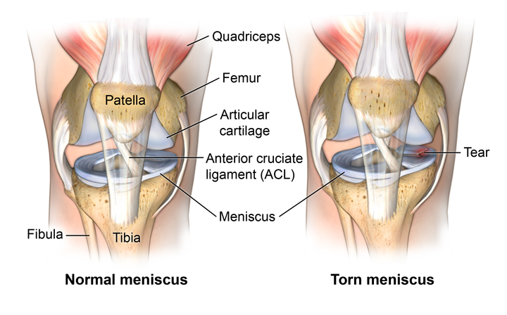
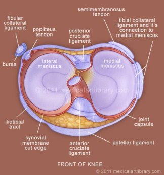
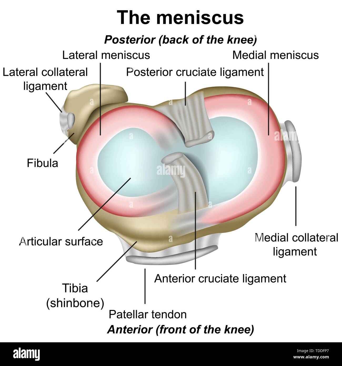
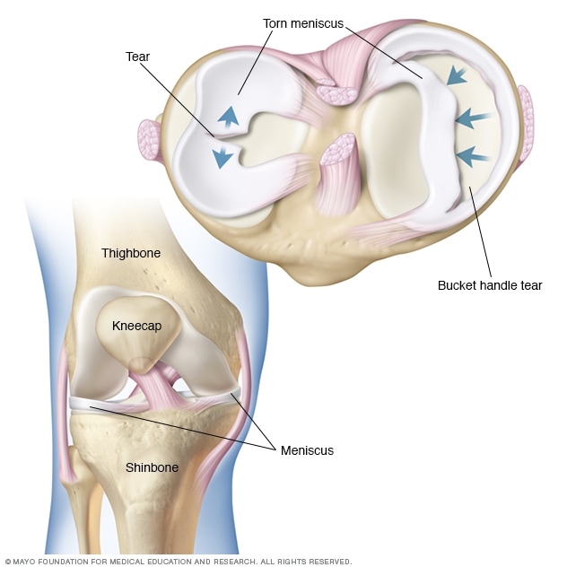
A torn meniscus is one of the most common knee injuries. Any activity that causes you to forcefully twist or rotate your knee, especially when putting your full weight on it, can lead to a torn meniscus. Each of your knees has two C-shaped pieces of cartilage that act like a cushion between your shinbone and your thighbone (menisci). A torn ... The menisci of the knee are two pads of fibrocartilaginous tissue which serve to disperse friction in the knee joint between the lower leg (tibia) and the thigh ...Latin: MenisciMeSH: D000072600Greek: μηνίσκος ("meniskos")Structure · Clinical significance We are pleased to provide you with the picture named Meniscus anatomy diagram.We hope this picture Meniscus anatomy diagram can help you study and research. for more anatomy content please follow us and visit our website: www.anatomynote.com. Anatomynote.com found Meniscus anatomy diagram from plenty of anatomical pictures on the internet.We think this is the most useful anatomy picture that ... Knee pain could be the result of a problem with any one of these components, or a combination of several. You may be experiencing knee pain and want to know the possible causes. The diagram, below, is a handy guide to the possible reasons for your pain. Pain at the front above the knee
What Is A Torn Meniscus
Diagram of Knee Joint showing Meniscus The meniscus is a crescent-shaped structure composed of cartilage that functions to distribute body weight evenly across the three bones that make up the knee joint: the thigh bone, shin bone, and knee cap.

Knee Joint Anatomy Movement Muscle Involvement How To Relief

Meniscus Tear

Lateral Meniscus Tear

Meniscus Knee Sports Orthobullets
/vector-illustration-of-a-meniscus-tear-and-surgery-871162428-03ac23d73f854954a8082f2ae3ce9219.jpg)
Meniscus Vs Cartilage Tear Of The Knee
Torn Meniscus Johns Hopkins Medicine
What Is The Meniscus And What Role Does It Play In The Knee Fit 2 Function Clinic
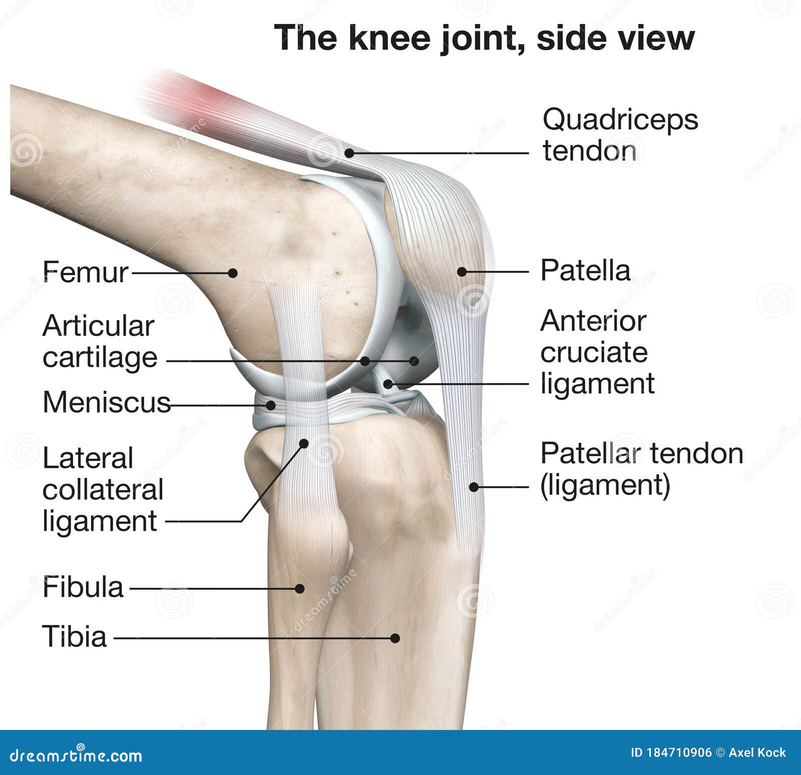
Knee Joint Anatomy Side View Medical 3d Illustration Stock Illustration Illustration Of Anatomy Tendon 184710906

Knee Joint Picture Image On Medicinenet Com

Torn Meniscus Anatomy And Causes Video Town Center Orthopaedic Associates
Meniscus Tear Orthopedics Medbullets Step 2 3
Meniscal Tears Jointsurgery In
Knee Pain Surgery Mcvay Physical Therapy
Knee And Meniscus Anatomy
The Knee Joint Articulations Movements Injuries Teachmeanatomy
3
Meniscal Tear Surgery Torn Meniscus Surgery Bayonne Bloomfield

The Knee Meniscus Injury

Meniscus Anatomy

Knee And Meniscus Anatomy Medical Vector Illustration Isolated On White Background Eps 10 Stock Vector Image Art Alamy
Meniscus Knees Knee Legs Skeleton Leg Thigh Joints Anatomy Bones Stock Vector Image Art Alamy
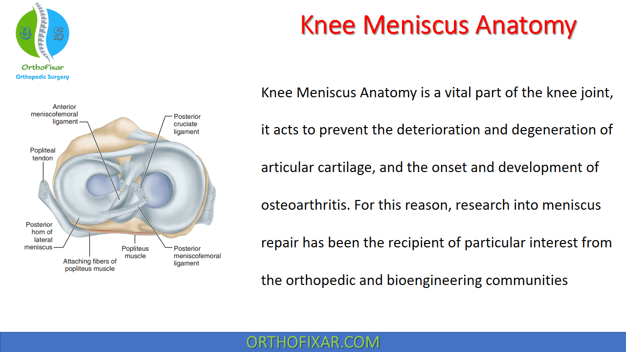
Knee Meniscus Anatomy Easy Explained Orthofixar 2021
Lateral Meniscus Physiopedia
Knee Meniscus Function Injuries Knee Pain Explained
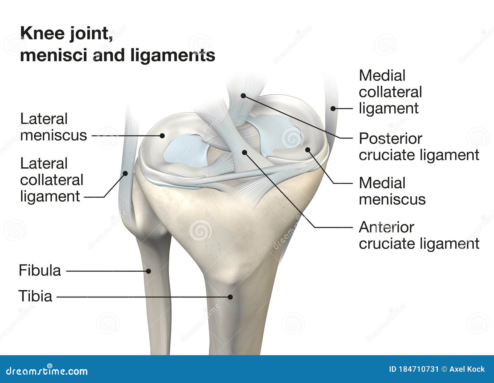
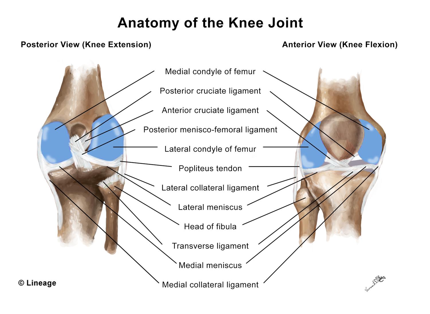
Knee Joint Anatomy Menisci And Ligaments Medically 3d Illustration Stock Illustration Illustration Of Joint Patella 184710731

Anatomy Of Knee

Knee Anatomy Creative Commons Diagram Radiology Case Radiopaedia Org
Knee Meniscus Anatomy
What Is A Meniscus With Pictures

A Anatomy Of The Meniscus Viewed From Above Adapted Image Reprinted Download Scientific Diagram

Understanding Meniscus Tears
Meniscus Knee Anatomy Medical Illustration Isolated On White Background Infographic Stock Vector Image Art Alamy

What Are The Parts Of The Knee Joint Systems4knees

Meniscal Injuries Page 1 Of 0 Sports Spinal Albury Primary Category Sports Spinal Albury
:max_bytes(150000):strip_icc()/WhatisMeniscalCyst_2549646_Final_1-dfb0f73f6d6a4548a7ef0485e7d7d869.jpg)
Meniscal Cysts Symptoms Causes Diagnosis And Treatment
Knee Anatomy Lyndon Bradley

Figure Anatomy Of The Right Knee Download Scientific Diagram
Meniscus Tears Orthoinfo Aaos
Torn Meniscus Symptoms And Causes Mayo Clinic
0 Response to "40 meniscus diagram of the knee"
Post a Comment