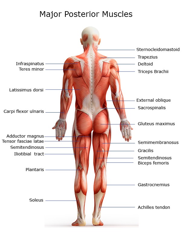
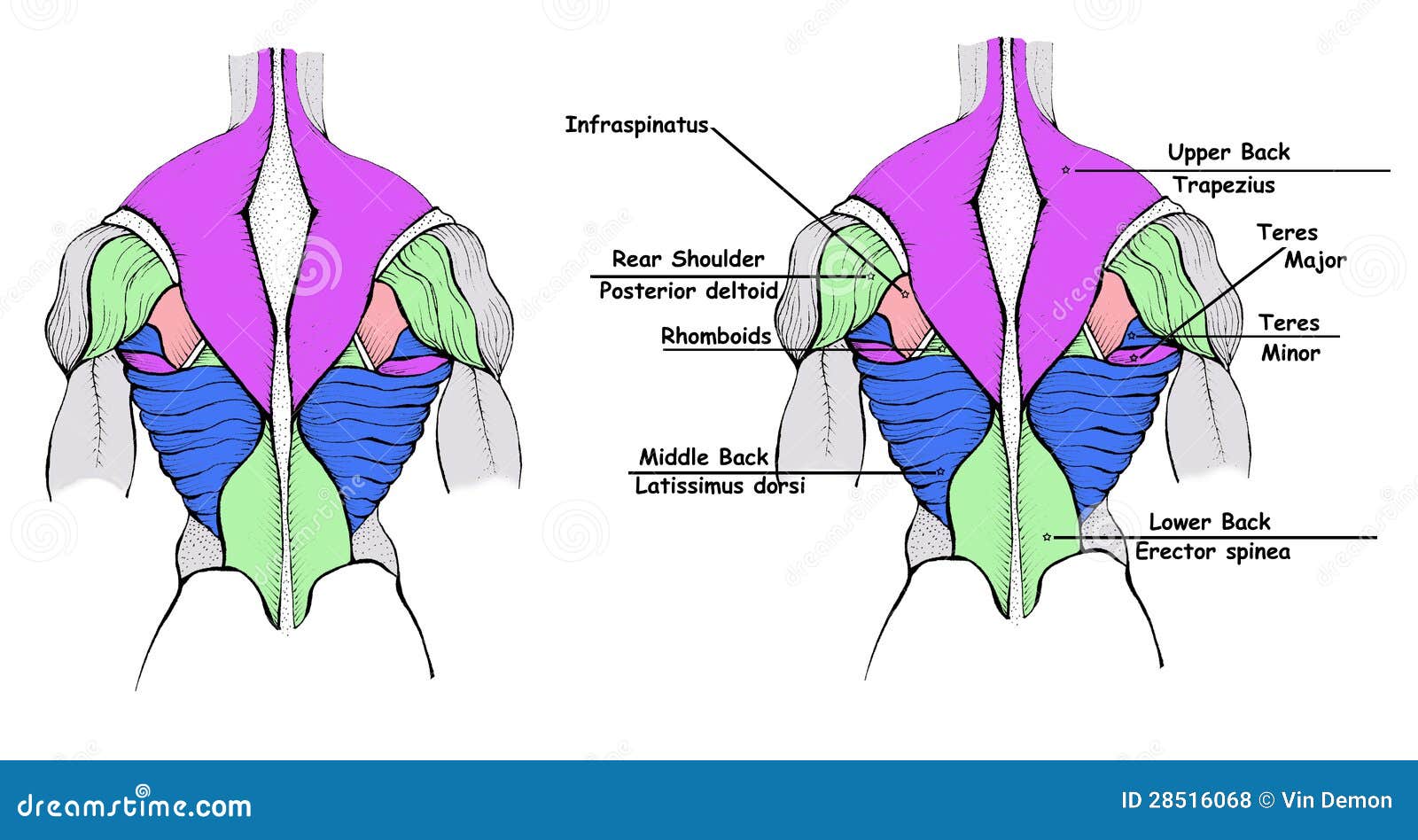
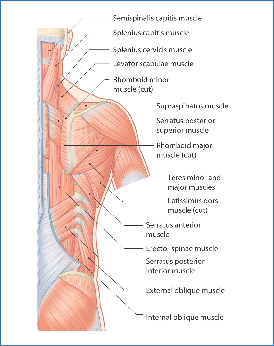
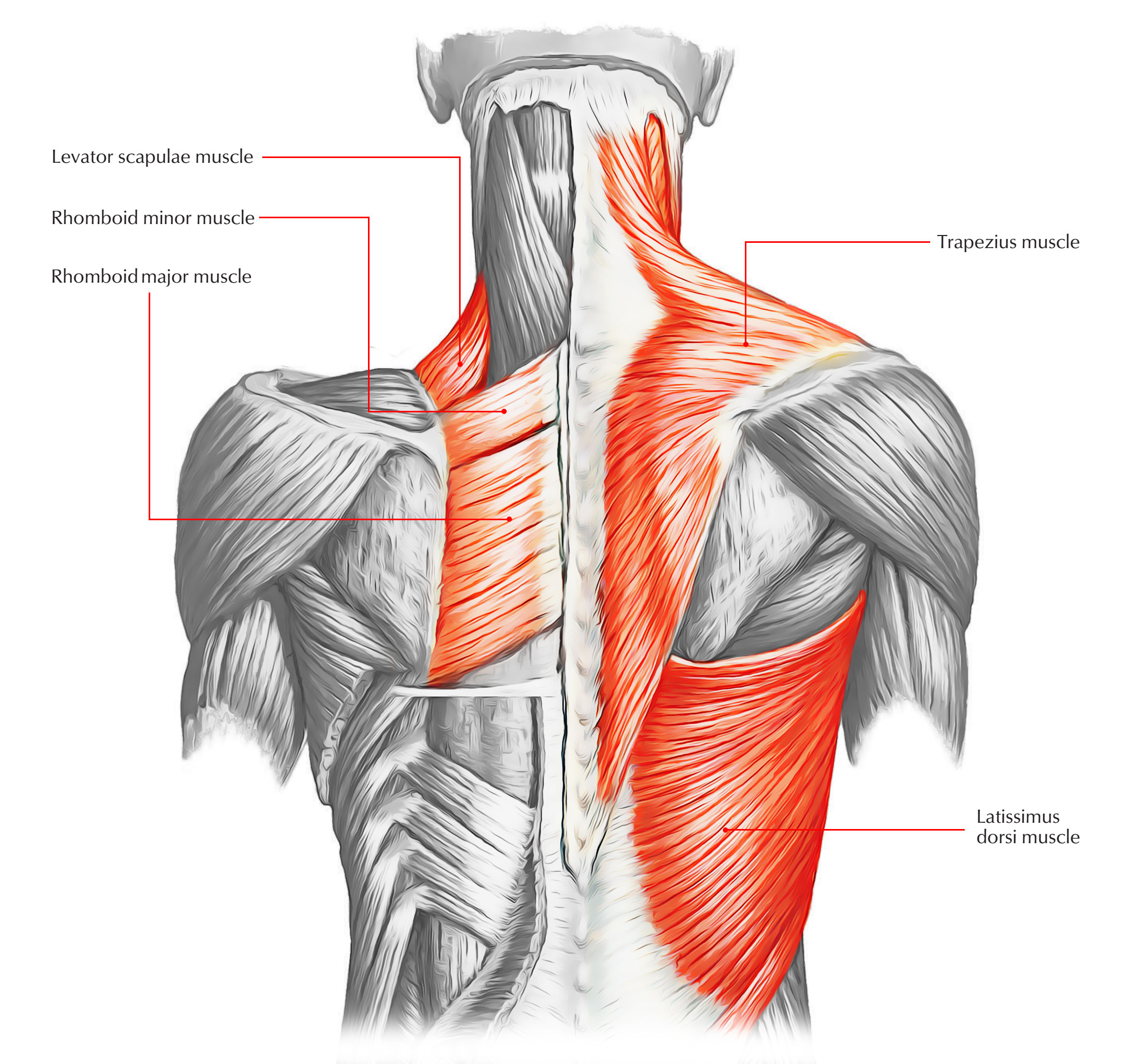
41 diagram of the back muscles
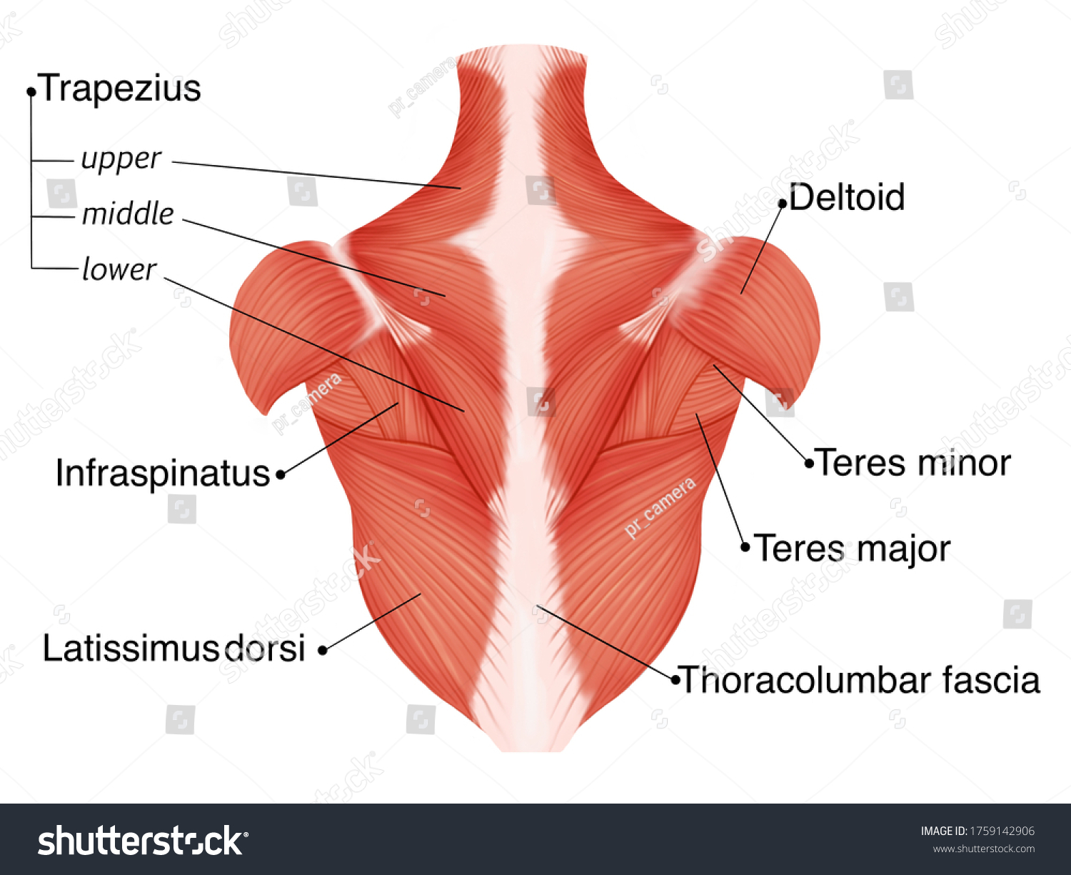
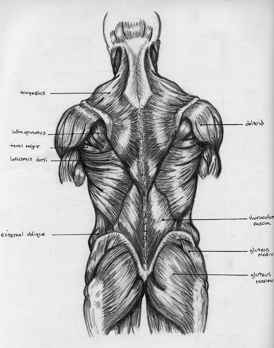
Upper Back Muscles - Medical Art Library The deltoid, teres major, teres minor, infraspinatus, supraspinatus (not shown) and subscapularis muscles (not shown) all extend from the scapula to the humerus and act on the shoulder joint. The trapezius and latissimus dorsi muscles connect the upper limb to the vertebral column. Both the deltoid and the trapezius are firmly attached to the spine of the scapula. Diagram Of Back Muscles Stock Photos, Pictures & Royalty ... Browse 561 diagram of back muscles stock photos and images available, or start a new search to explore more stock photos and images. Newest results Male fitness model Anatomy engraving Male and female body chart Male Human Body Muscle map leg back muscles 3d medical vector illustration on white... Human Muscle
Human Anatomy - Back View of Muscles - Logo of the BBC Human Anatomy - Back View of Muscles Click on the labels below to find out more about your muscles. More human anatomy diagrams: front view of muscles, skeleton, organs, nervous system Flex some...
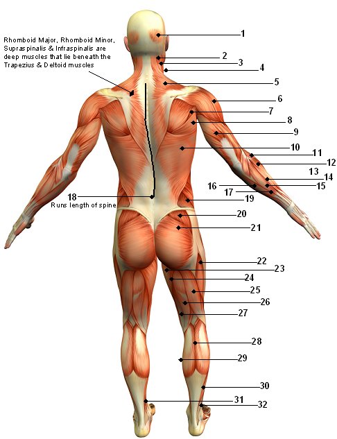
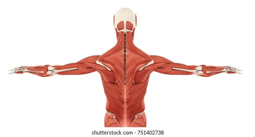
Diagram of the back muscles
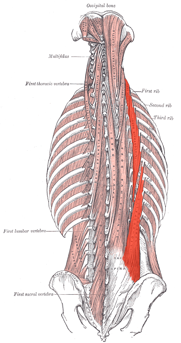
Back muscles: Anatomy and functions | Kenhub The transversospinal muscles gather three groups of back muscles: The semispinalis muscle, which is topographically divided into the semispinalis capitis, semispinalis cervicis and semispinalis thoracis. They span between the transverse and spinous processes of the regional vertebrae. Back Muscles: Names And Diagram - Science Trends Muscles found in the deep group include the spinotransversales, erector spinae (composed of the iliocostalis, longissimus, and spinalis), the transversospinales, and the segmental muscles. "The best way to strengthen back muscles is in a static position. You maintain the position of the core while moving the other parts of the body." Human Muscles Diagram Front And Back at Anatomy This is a diagram of the larger and more surface muscles of the low back. Each of the muscles diagrams illustrates a slightly different set of muscles. Back muscle diagram with lower back anatomy. The extrinsic back muscles are located in the back, but act to produce movements of the shoulder and assist respiration.
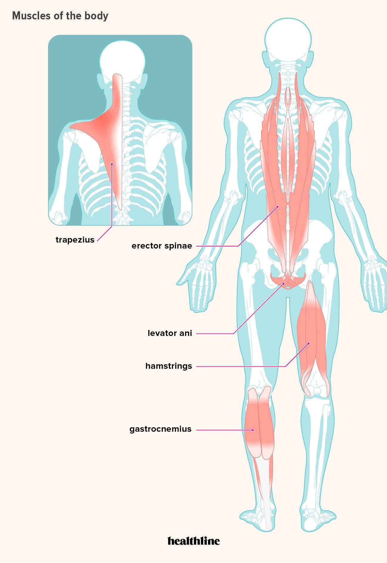
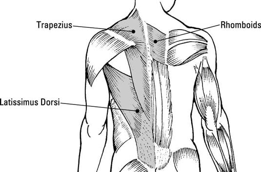
Diagram of the back muscles. Back Muscles: Anatomy, Function, Treatment The muscles in the back are the trapezius, rhomboids, latissimus dorsi, erector spinae, multifidus, and quadratus lumborum. How can I prevent back pain? Keep your back muscles in good shape to prevent back pain. Exercises that strengthen the core (abdominals and lower back) can help to protect the spine from damage. Muscles of the low back - Learn Muscles Muscles of the Low Back and Pelvis - posterior superficial view Muscles of the Low Back and Pelvis - right lateral deep view Muscles of the Low Back and Pelvis - right lateral superficial view Upper Back Muscles Diagram | Quizlet Start studying Upper Back Muscles. Learn vocabulary, terms, and more with flashcards, games, and other study tools. Back Pain Symptoms Chart - THC Bone and Joint Back Pain Symptoms Chart. The vast majority of back problems improve on their own or with nonsurgical treatment. There are a few warning signs, however, that may indicate serious spinal problems. If you experience any of these symptoms, seek medical attention immediately. Loss of control of the bowel or bladder and retention of urine may ...
Muscles Body Diagram Stock Illustrations - 571 Muscles ... Muscle chart - GERMAN LABELING - most important muscles of the human body - colored front and back view - isolated vector Vector illustration about how muscles grow. Medical educational diagram and scheme with satellite cell and fusion of cells. Lower Back Pain - An Overview of the Key Muscles Involved ... Gluteus Medius. This muscle is a major generator of lower back and hip pain, as well as being responsible for complaints of a burning sensation along the posterior superior iliac spine (PSIS) and sacroiliac joint. Pain is often mistaken for lumbago- type pain, with discomfort (such as tenderness) into the buttocks and superior thigh. Back Muscles, Back Muscle Diagram - Muscleblitz.com The following diagram shows all the major back muscles back muscles Back Exercises How to build a better back The top three back training mistakes to avoid The forgotten traps exercise build incredible thickness The deadlift back killer or mass builder How To Build A Wide Back Back muscle anatomy Images, Stock Photos & Vectors ... Back muscle anatomy images. 26,354 back muscle anatomy stock photos, vectors, and illustrations are available royalty-free. See back muscle anatomy stock video clips. Image type.
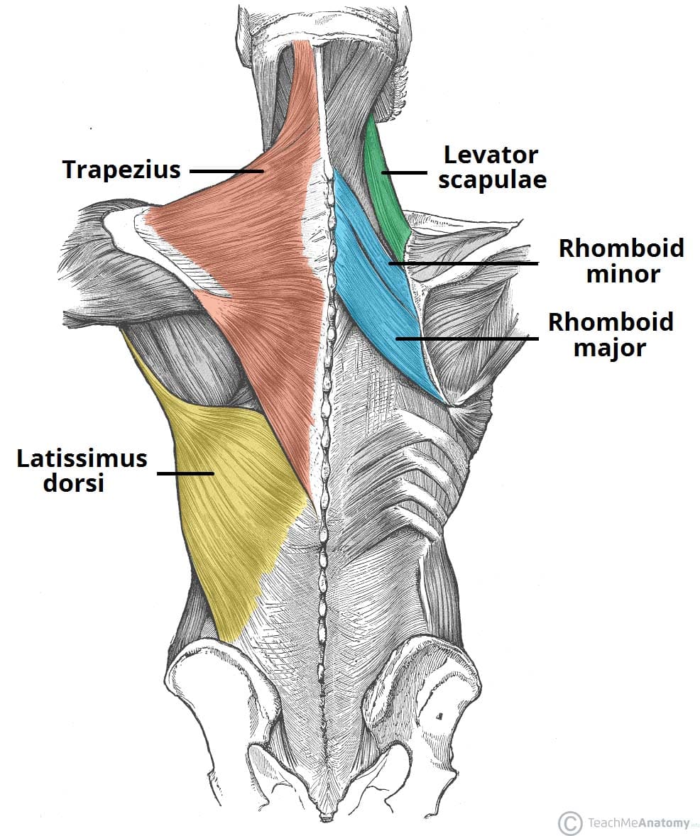
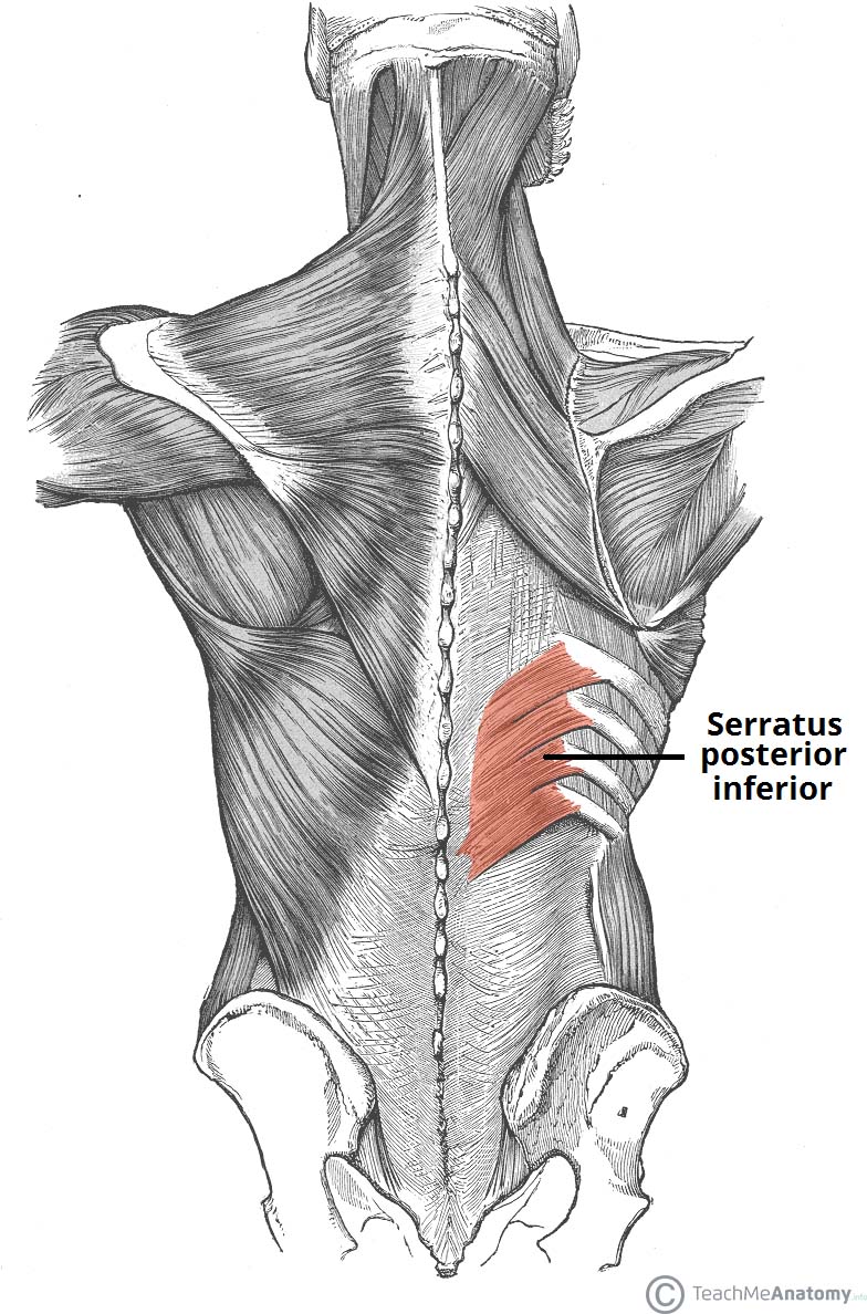
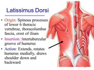
Back Muscles Diagram Stock Photos, Pictures & Royalty-Free ... Labeled human anatomy diagram of man's arm, shoulder and upper back muscles in a posterior view on a white background. Exercise Step with Reverse Crunch by healthy woman. Rectus Abdominis - Abdominal Muscles - Anatomy Muscles isolated Correct alignment of human body in standing posture Back Muscles: Anatomy of Upper, Middle & Lower Back Pain ... There are five pairs of back muscles that help move the shoulders and upper arms. They are the latissimus dorsi, supraspinatus, infraspinatus, teres major, and teres minor. Latissimus Dorsi The latissimus dorsi, often called the lats, are large wing-shaped muscles that extend from the upper to the lower back. They help move the arms and shoulders. Shoulder Muscles Anatomy, Diagram & Function | Body Maps Supraspinatus: This small muscle is located at the top of the shoulder and helps raise the arm away from the body. Four muscles—the supraspinatus, infraspinatus, teres minor, and subscapularis—make... Anatomy of the back: Spine and back muscles | Kenhub The superficial back muscles include the trapezius, latissimus dorsi, levator scapulae, rhomboids and serratus posterior muscles . Let us introduce you to each of these muscles presented in our diagram. Trapezius The trapezius muscle consists of three parts; descending, transverse and ascending.
Anatomy of the spine and back - e-Anatomy - IMAIOS The muscles of the back with the surface (trapezius, latissimus dorsi, thoracolumbar fascia, deltoid) and intermediate layers (serrated muscles, external and internal oblique muscle). The back muscles represented on an anatomical chart and on a schematic view of the origin and insertion of the proper muscles of the back (iliocostal muscle of ...
Back anatomy: Diagram and overview - Medical News Today There are three different groups of muscles in the back. These are called the superficial, intermediate, and intrinsic muscles. The sections below cover these in more detail. Superficial muscles...
Shoulder Pain Diagram: Diagnosis Chart - Shoulder Pain Exp Our next shoulder pain diagram focuses on problems around the back of the shoulders and the shoulder blades. There is some overlap with some of the conditions we've already looked at the can refer pain down the side and back of the arms as well as the front. A. Muscle Strain. Straining the upper trapezius muscles is a common cause of pain across the top part of the back of the …
Back Muscles Diagram | Quizlet Back Muscles Diagram | Quizlet. Upgrade to remove ads. Only $2.99/month.
Muscular System Diagram Posterior (Back) View - Sport ... by Sport Fitness Advisor Staff This muscular system diagram shows the major muscle groups from the back or posterior view. To see a muscular system picture from the anterior (front) view click here. Occipitalis Semispinalis Capitis Splenius Capitis Sternocleidomastoid Trapezius Deltiod Teres Minor Teres Major Triceps Brachii Latissimus Dorsi
17 Back Muscles That Cause the MOST Back Pain (and how to ... Bonus Content: The 7-Day Back Pain Cure Back pain is one of the top reasons for missed work and second only to upper-respiratory infections for causing doctor visits. Most of the time, back muscle pain is diagnosed then "treated" with little more than a prescription of rest, painkillers and muscle relaxants.
Anatomy Of Back Muscles Diagram Anatomynote.com found Anatomy Of Back Muscles Diagram from plenty of anatomical pictures on the internet. We think this is the most useful anatomy picture that you need. You can click the image to magnify if you cannot see clearly. This image added by admin. Thank you for visit anatomynote.com.
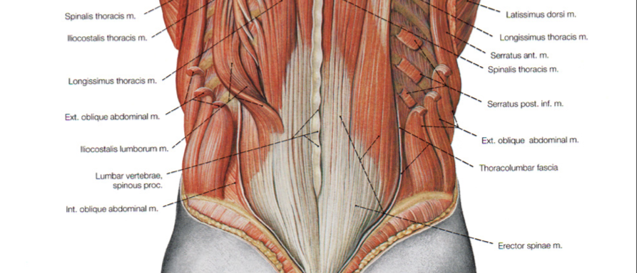
Lumbar Spine Anatomy, Diagram & Function | Body Maps Muscles connect to the vertebrae and bones via ligaments, flexible bands of fibrous tissue. The deep muscles of the back fit into or affix parts of themselves to the grooves in the spinous...
Muscles of the Back - TeachMeAnatomy The muscles of the back can be arranged into 3 categories based on their location: superficial back muscles, intermediate back muscles and intrinsic back muscles.The intrinsic muscles are named as such because their embryological development begins in the back, oppose to the superficial and intermediate back muscles which develop elsewhere and are therefore classed as extrinsic muscles.
Lower Back Muscle Anatomy and Low Back Pain However, there are many back muscles which can cause pain. Please refer to the Lower Back Muscle picture below to see all of the muscles of the back. The pelvic floor muscles also help increase this pressure, which provides stability to the spine and trunk. Common hip and back pain causes include injury to muscles from overuse, disc injury/degeneration, or spinal stenosis. To …
Human Muscles Diagram Front And Back at Anatomy This is a diagram of the larger and more surface muscles of the low back. Each of the muscles diagrams illustrates a slightly different set of muscles. Back muscle diagram with lower back anatomy. The extrinsic back muscles are located in the back, but act to produce movements of the shoulder and assist respiration.
Back Muscles: Names And Diagram - Science Trends Muscles found in the deep group include the spinotransversales, erector spinae (composed of the iliocostalis, longissimus, and spinalis), the transversospinales, and the segmental muscles. "The best way to strengthen back muscles is in a static position. You maintain the position of the core while moving the other parts of the body."
Back muscles: Anatomy and functions | Kenhub The transversospinal muscles gather three groups of back muscles: The semispinalis muscle, which is topographically divided into the semispinalis capitis, semispinalis cervicis and semispinalis thoracis. They span between the transverse and spinous processes of the regional vertebrae.



:background_color(FFFFFF):format(jpeg)/images/library/12063/primary-authochtone-back-muscles_english.jpg)



:background_color(FFFFFF):format(jpeg)/images/library/14074/Back_muscles.png)



:watermark(/images/watermark_5000_10percent.png,0,0,0):watermark(/images/logo_url.png,-10,-10,0):format(jpeg)/images/overview_image/2247/O4LWJymz1b1eMKEx25fWw_superficial-intermediate-deep-back-muscles_english.jpg)

/GettyImages-56372373-1bb8a07f23d84f439c3e80ecf3c472d2.jpg)


0 Response to "41 diagram of the back muscles"
Post a Comment