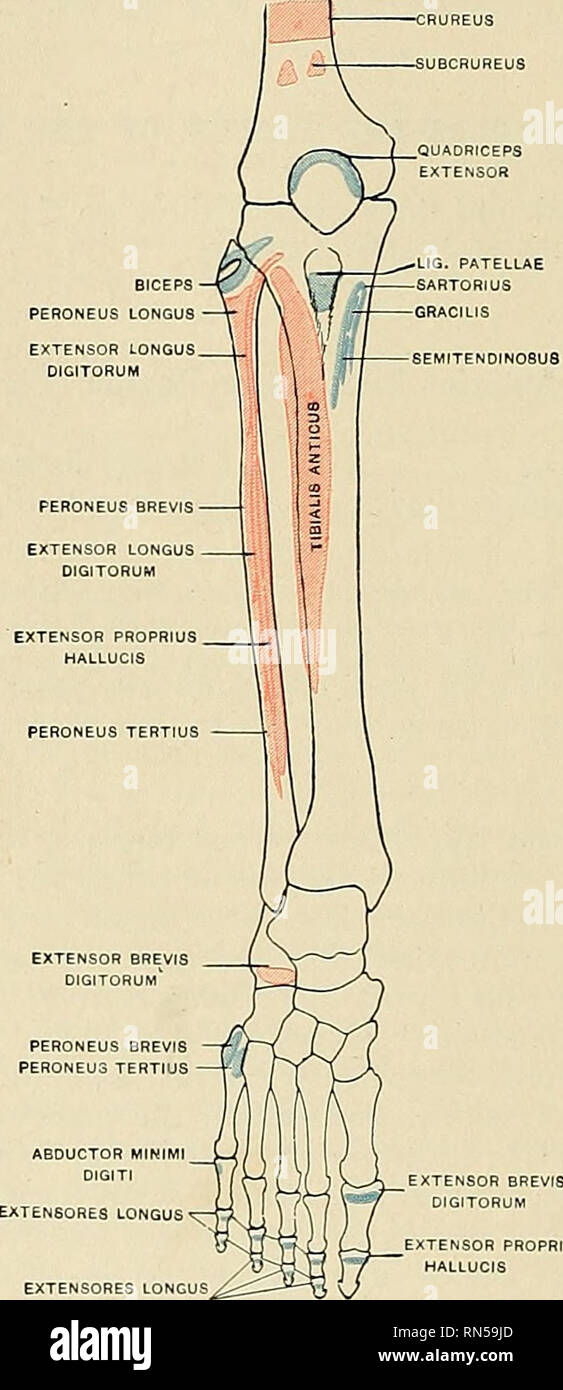
38 ankle muscles and tendons diagram
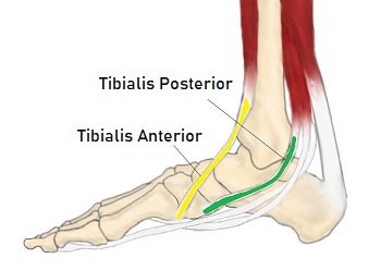
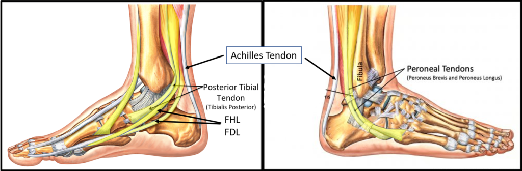
Tibialis posterior muscle - Wikipedia Structure. The tibialis posterior muscle originates on the inner posterior border of the fibula laterally. It is also attached to the interosseous membrane medially, which attaches to the tibia and fibula.. The tendon of the tibialis posterior muscle (sometimes called the posterior tibial tendon) descends posterior to the medial malleolus. It terminates by dividing into plantar, main, … Hip Anatomy, Pictures, Function, Problems & Treatment 28-07-2010 · Hip Anatomy, Function and Common Problems Front View of the Hip Joint Bones. Normally, a smooth cushion of shiny white hyaline (or articular) cartilage about 1/4 inch thick covers the femoral head and the acetabulum.The articular cartilage is kept slick by fluid made in the synovial membrane (joint lining).
Skeletal System: Anatomy and Function, Diagram, Diseases ... 30-08-2018 · The skeletal system is the foundation of your body, giving it structure and allowing for movement. We’ll go over the function and anatomy of …
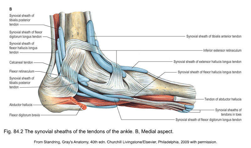
Ankle muscles and tendons diagram
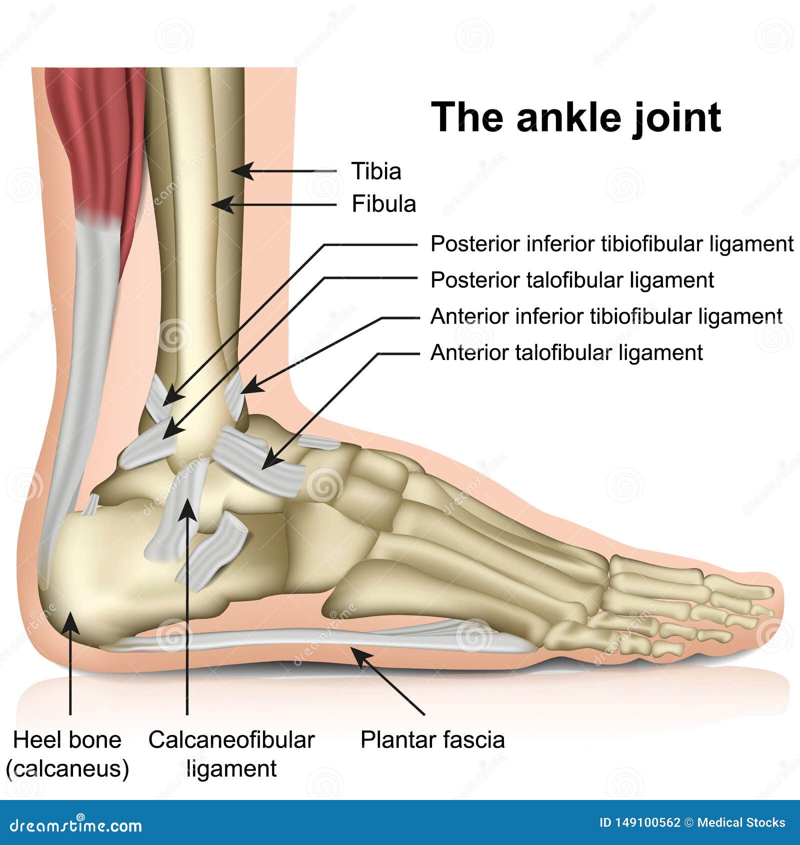
Ankle - Wikipedia The ankle, or the talocrural region, or the jumping bone (informal) is the area where the foot and the leg meet. The ankle includes three joints: the ankle joint proper or talocrural joint, the subtalar joint, and the inferior tibiofibular joint. The movements produced at this joint are dorsiflexion and plantarflexion of the foot. In common usage, the term ankle refers exclusively to the ankle ... Chart of Major Muscles on the Front of the Body with Labels 21-06-2012 · We have a lot of muscles in our bodies (literally, over 600). Muscles allow us to move and function. In general, they work in pairs. Usually as one muscle contracts (or shortens), the opposing muscle (known as the antagonist) elongates and vice versa.For example, think about when you bend your arm to bring food to your mouth. Foot (Anatomy): Bones, Ligaments, Muscles, Tendons, Arches ... 25-07-2017 · Foot Definition. The foot is a part of vertebrate anatomy which serves the purpose of supporting the animal’s weight and allowing for locomotion on land. In humans, the foot is one of the most complex structures in the body. It is made up of over 100 moving parts – bones, muscles, tendons, and ligaments designed to allow the foot to balance the body’s weight on …
Ankle muscles and tendons diagram. Nervous System: Anatomy and Function - PT Direct The tension in the tendons and muscles is constantly monitored and if pain increases the brain gets the message and I may choose to stop and stretch. Worked example; learning to squat. What about a movement for which the brain doesn’t have an established motor pattern to recall, such as learning to do a squat holding dumbbells for the first time. NASM Study Guide 7th Ed 2022 - Pass the NASM exam for FREE! 15-09-2021 · We promise all the tools to help you become a successful NASM Certified Personal Trainer. Be sure to Bookmark! Lastly, this page is crafted for the layout of the NASM 7th Edition text, if you would like the 6th Edition layout, check it out here.. Don’t forget to download our 14 step NASM CPT exam preparation checklist to ensure that you pass the test. Biceps Tendon Tear at the Shoulder - OrthoInfo - AAOS Tendons attach muscles to bones. Your biceps tendons attach the biceps muscle to bones in the shoulder and elbow. If you tear the biceps tendon at the shoulder, you may lose some strength in your arm and have pain when you forcefully turn your arm from palm down to palm up. Pulled Muscles, Muscle Strain and Scar Tissue Treatment 05-03-2019 · The diagram below is a comparison of the same injury treated ... In this position you can push against the wall with your foot and at the same time keep your ankle joint from moving. The muscles contract but the ankle ... Without this information the muscles, tendons and ligaments are constantly second-guessing the position of ...
Foot (Anatomy): Bones, Ligaments, Muscles, Tendons, Arches ... 25-07-2017 · Foot Definition. The foot is a part of vertebrate anatomy which serves the purpose of supporting the animal’s weight and allowing for locomotion on land. In humans, the foot is one of the most complex structures in the body. It is made up of over 100 moving parts – bones, muscles, tendons, and ligaments designed to allow the foot to balance the body’s weight on … Chart of Major Muscles on the Front of the Body with Labels 21-06-2012 · We have a lot of muscles in our bodies (literally, over 600). Muscles allow us to move and function. In general, they work in pairs. Usually as one muscle contracts (or shortens), the opposing muscle (known as the antagonist) elongates and vice versa.For example, think about when you bend your arm to bring food to your mouth. Ankle - Wikipedia The ankle, or the talocrural region, or the jumping bone (informal) is the area where the foot and the leg meet. The ankle includes three joints: the ankle joint proper or talocrural joint, the subtalar joint, and the inferior tibiofibular joint. The movements produced at this joint are dorsiflexion and plantarflexion of the foot. In common usage, the term ankle refers exclusively to the ankle ...
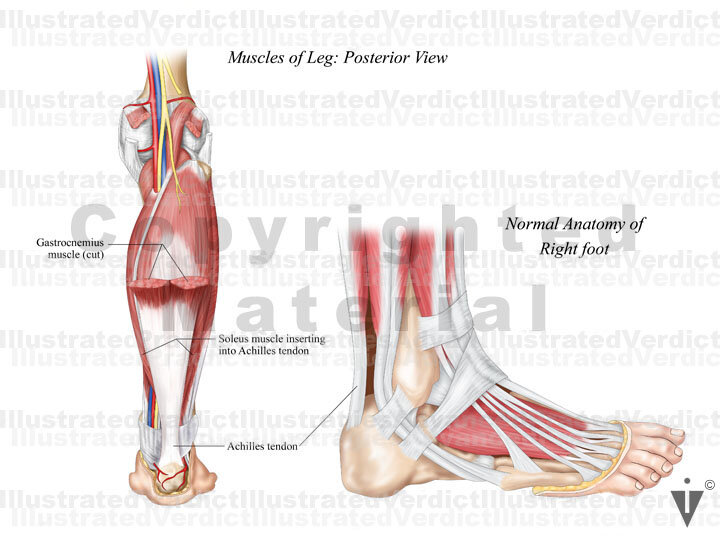
Stock Ankle: Normal Anatomy — Illustrated Verdict
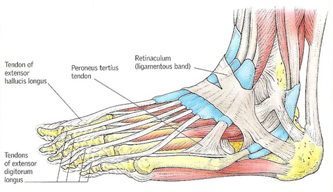
Foot & Ankle Tendons: Anatomy, Function & Injuries

BeneFIT PT's Anatomy Series: The Ankle

ANKLE/FOOT ANATOMY Diagram | Quizlet

Tibialis Posterior: Pain On The Inside Of The Ankle - Pure ...
Ankle Anatomy - Be In Motion Physiotherapy

11.2a Muscles & Movement | Grace's Blog
MUSCLES OF THE ANKLE AND FOOT

Why Shape Matters — Boot Solutions Japan
Patient Education | Concord Orthopaedics

Hand & Wrist Pain | Stem Cell Therapy | KOPI | Kingston

Anatomy of the lateral ankle (reproduced with permission from ...

The leg, ankle, and foot - Knowledge @ AMBOSS

Ankle Posterolateral Approach - Approaches - Orthobullets
Applied anatomy of the lower leg, ankle and foot
Anatomy, descriptive and applied. Anatomy. 526 THE MUSCLES ...

Muscles of the lower leg and foot | Human Anatomy and ...

Foot and Ankle | Musculoskeletal Key

A Patient's Guide to Foot Anatomy | 2020 OrthoNorCal, Los ...

Ligaments In The Foot - JOI Jacksonville Orthopaedic Institute

Types of Tendon Surgery | Foot & Ankle Associates of Florida

Basic Foot and Ankle Anatomy - Neural and Vascular - Physiopedia
The Ankle Joint, Tendons of the Ankle Joint Foot Anatomy ...

Human leg muscle illustration, Foot Anatomy Muscle Ankle Bone ...
foot (anatomy)

Anatomy of the Foot and Ankle | OrthoPaedia
Ankle Tendon Tear - Centeno-Schultz Clinic
Ankle and foot anatomy: Bones, joints, muscles | Kenhub

Foot Ankle Tendons Ligaments Joints Bones Stock Illustration ...
Ankle Anatomy - Be In Motion Physiotherapy

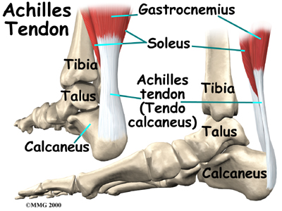
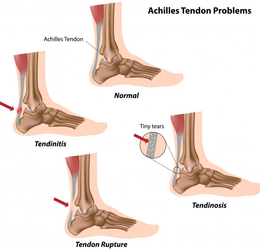
Why is the prototypical patient with a ruptured Achilles ...
What is the Anatomy of the Ankle? (with pictures)
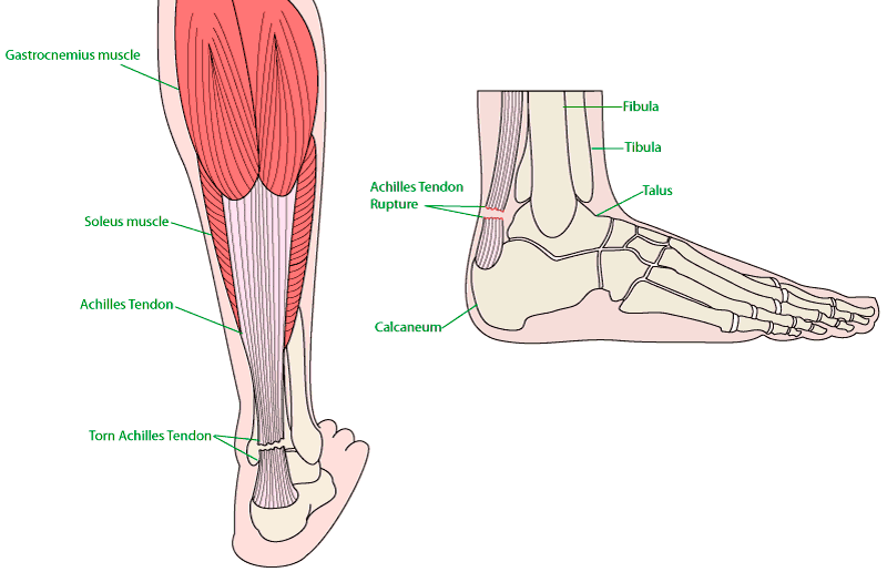
Achilles Tendon Rupture - Orthoanswer

BeneFIT PT's Anatomy Series: The Ankle
Ankle Joint: Anatomy | Concise Medical Knowledge
Ankle and foot anatomy: Bones, joints, muscles | Kenhub

Ankle Sprains: Symptoms, Treatments & Prevention | Watsonia ...

Basic Ankle Anatomy | Joint Pain Info
0 Response to "38 ankle muscles and tendons diagram"
Post a Comment