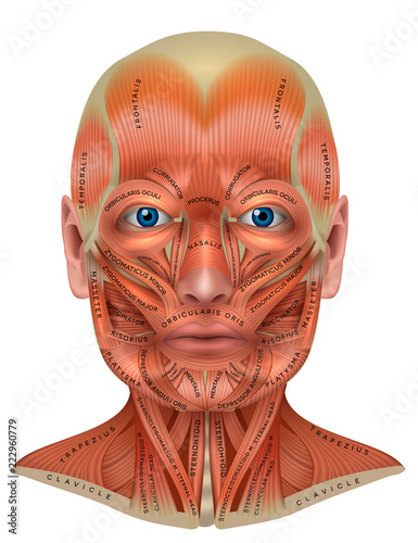
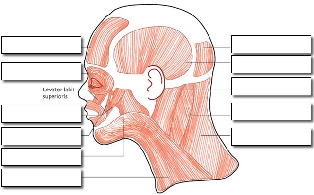
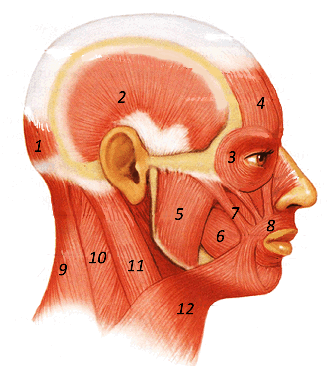
8 head and neck muscle diagram
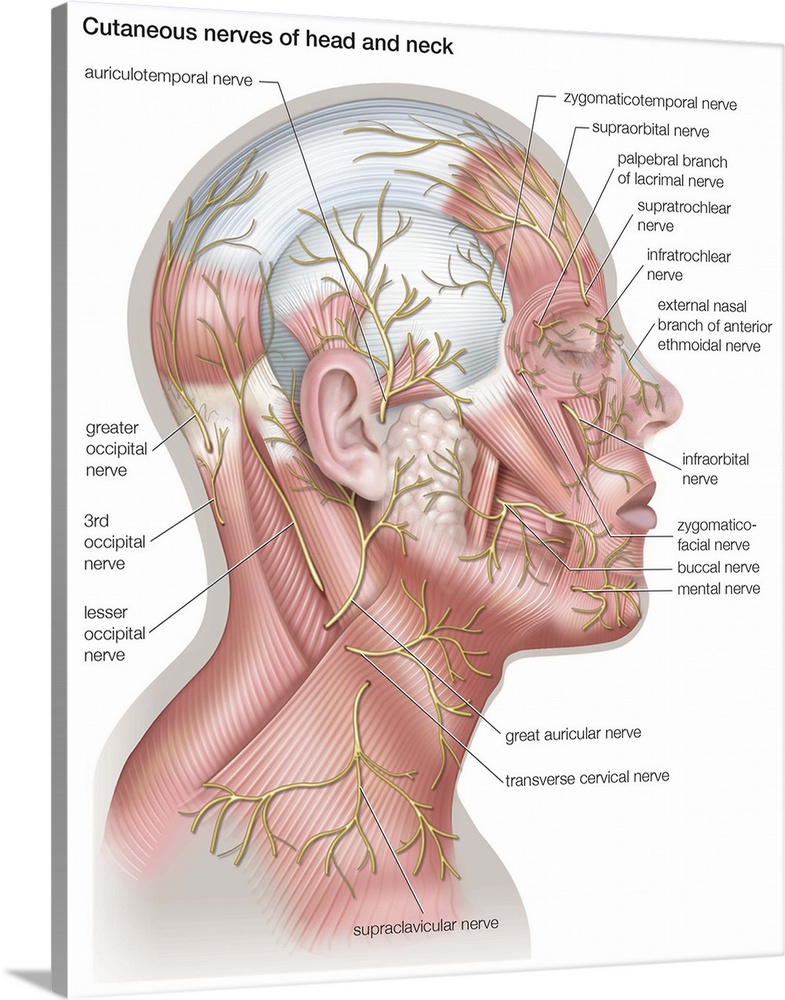
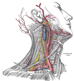
Head and neck anatomy,muscles,blood supply diagram. This diagram depicts throat and neck anatomy. Surface anatomy and surface markings bibliographic record list of illustrations the external jugular vein varies in size, bearing an inverse proportion to the other veins of the neck, it is. Some of the axial muscles may seem to blur the boundaries because they cross over to the appendicular skeleton. The first grouping of the axial muscles you will review includes the muscles of the head and neck, then you will review the muscles of the vertebral column, and finally you will review ...
Body System ( T022 ) SnomedCT. 281232002. English. Vascular structure head & neck, Vascular structure of head and neck, Vascular structure of head and neck (body structure) Spanish. estructura vascular de cabeza y cuello (estructura corporal), estructura vascular de cabeza y cuello. Derived from the NIH UMLS ( Unified Medical Language System )
:background_color(FFFFFF):format(jpeg)/images/library/10667/image2.png)
Head and neck muscle diagram
Here are a number of highest rated Posterior Neck Anatomy Diagram pictures upon internet. We identified it from reliable source. Its submitted by presidency in the best field. We agree to this kind of Posterior Neck Anatomy Diagram graphic could possibly be the most trending topic following we ration it in google lead or facebook. Nicole Long Date: January 29, 2022 A diagram showing nerves in the head and neck.. Nerves in the neck, medically referred to as the cervical spine, help transmit information along the pathways of the central and peripheral nervous system, including sensory and motor skills processes.The cervical spine consists of eight different sets of nerves. CT scan of head and neck : Radiological anatomy of the head and neck on a CT in axial, coronal, and sagittal sections, and on a 3D images
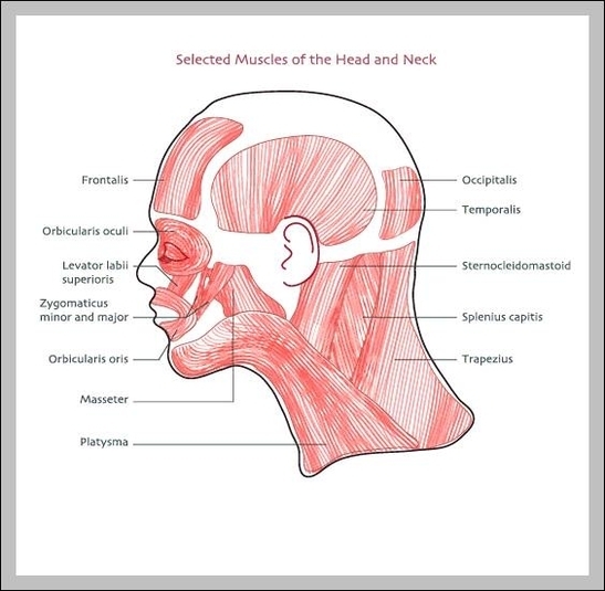
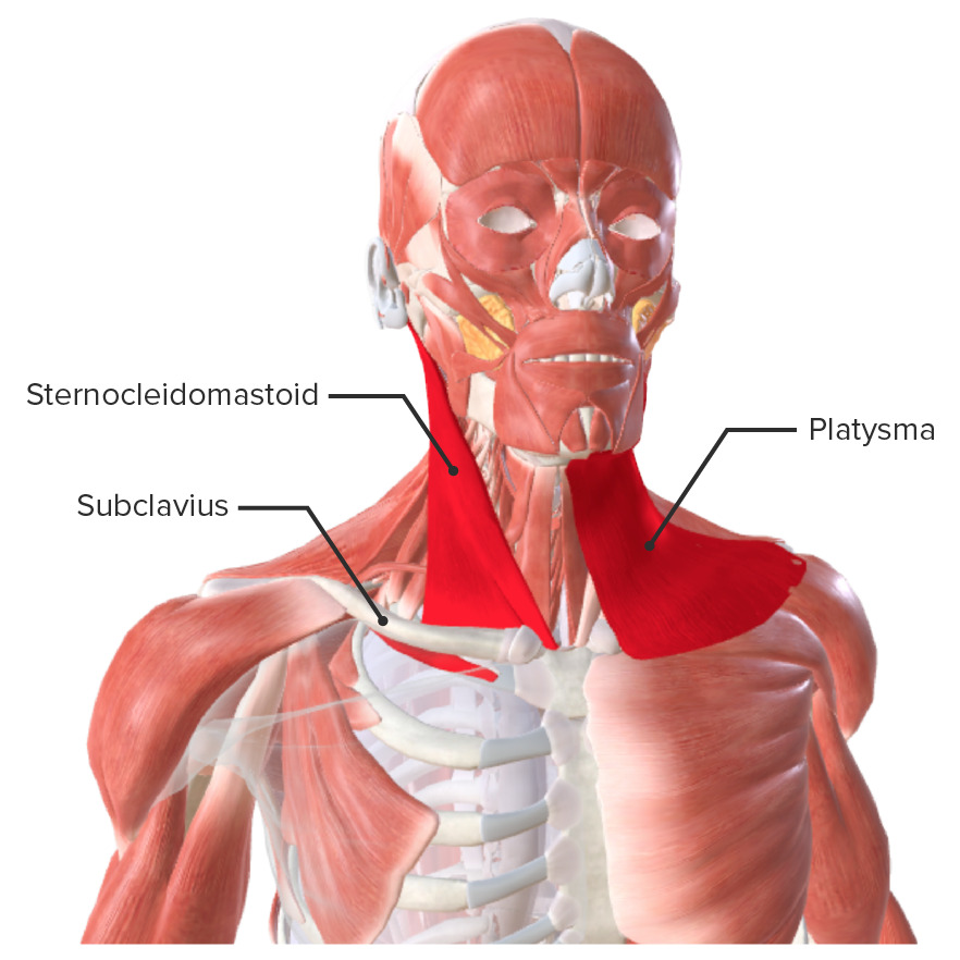
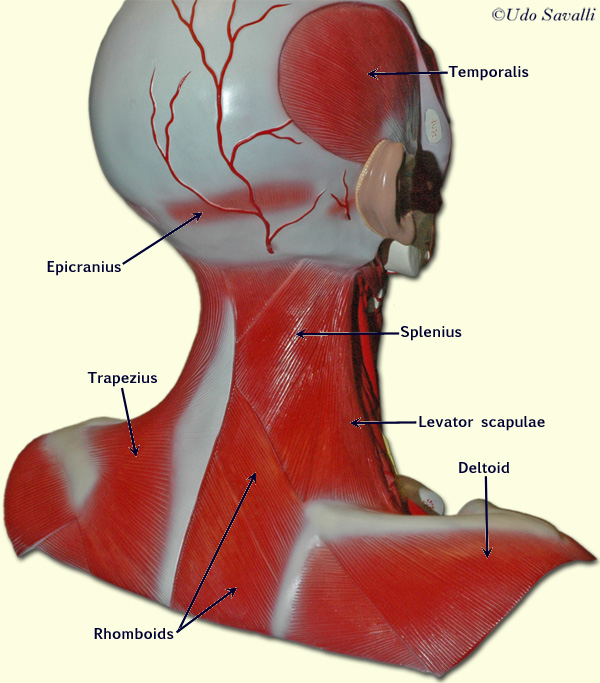
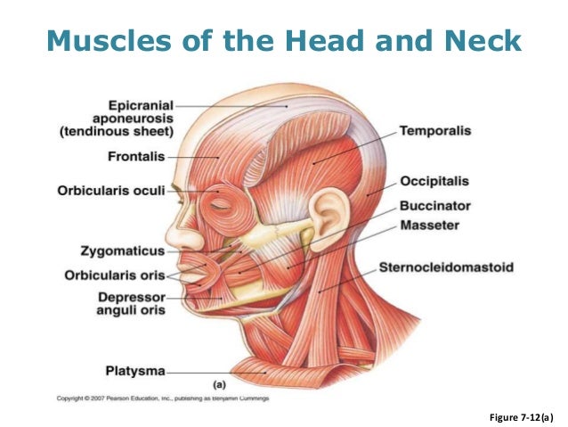
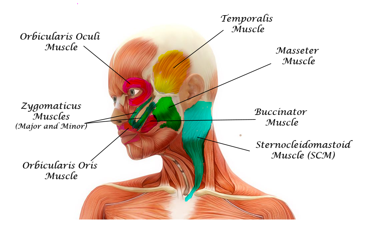
Head and neck muscle diagram. The neck is the bridge between the head and the rest of the body. It is located in between the mandible and the clavicle, connecting the head directly to the torso, and contains numerous vital structures. It contains some of the most complex and intricate anatomy in the body and is comprised of numerous organs and tissues with essential structure and function for normal physiology. Muscles of the Head and Neck - Chart for Massage Therapists. Assists in depressing mandible and tightens the fascia of neck depresses lower lip. Brachial plexus and subclavian artery pass between the middle and anterior scalene. Palpate between trapezius and SCM above levator scapula.Wrap around deeper neck muscles. A single platysma muscle is only shown in the lateral view of the head muscles in Figure 8-4. There are two platysma muscles, one on each side of the neck. Each is a broad sheet of a muscle that covers most of the anterior neck on that side of the body. The other anterior neck muscles are below ... Diagrams of muscles of the head, neck and intrinsic muscles of the back. All image files licensed under creative commons by 3.0 openstax . Muscle, origin, insertion, action, innervation, artery, notes, image. Splenius capitis and splenius cervicis: Homologies, evolution, and proposal of a mammalian and a veterinary muscle ontology. Head And Neck Anatomy Posterior …
The Muscles of Facial Expression. View Article. The Muscles of Mastication. View Article. The Tongue. View Article. Anatomy Video Lectures ... Cross-sectional labeled anatomy of the head and neck of the domestic cat on CT imaging (bones of the skull, cervical spine, mandible, hyoid bone, muscles of the neck, nasal cavity and paranasal sinuses, oral cavity, larynx) This diagram depicts the buccinator muscle in relation to the maxible, mandible, orbicularis oris ... Muscles of the oral group play key roles in respiration, ... 03/11/2021 · Head and neck; Trunk wall; Each chart groups the muscles of that region into its component groups, making your revision a million times easier. For example, upper limb muscles are grouped by shoulder and arm, forearm and hand. Next to each muscle, you’ll find its origin(s), insertion(s), innervation(s) and function(s). Muscle origins and insertions. Many muscles are …
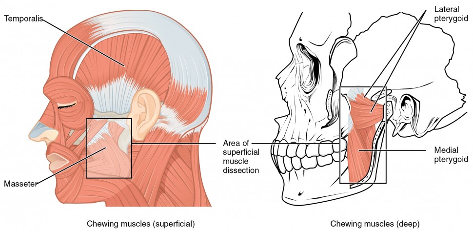
Mary Elizabeth Date: January 27, 2022 An anatomical illustration showing many of the muscles in the neck.. The neck muscles provide support for the head and allow the neck to move. Neck exercises are those exercises that involve the muscles of the neck, of which there are many, including the deltoid muscle, the trapezius muscle, the scalene muscles, the sternocleidomastoid muscle, the levator ... The primary muscles of mastication (chewing food) are the temporalis, medial pterygoid, lateral pterygoid, and masseter muscles. The four main muscles of mastication attach to the rami of the mandible and function to move the jaw (mandible). The cardinal mandibular movements of mastication are elevation, depression, protrusion, retraction, and side to side movement. To augment the process of ... Educational video describing the muscle anatomy of the neck.Become a friend on facebook:http://www.facebook.com/drebraheimFollow me on twitter:https://twitte The muscles in the human body's head and neck control facial expressions, enable the mouth to talk and chew, and allow the head to move. Explore the anatomy of these muscles, and understand how ...
Feb 22, 2012 - This Pin was discovered by M@ryam Ahmadi. Discover (and save!) your own Pins on Pinterest
July 6, 2020 - Nasalis: This muscle helps you scrunch your nose by compressing the bridge of the nose and pulling the nostrils open. Mentalis: This muscle causes wrinkles in your chin. Sternocleidomastoid: This large neck muscle helps rotate the head upward and side to side.
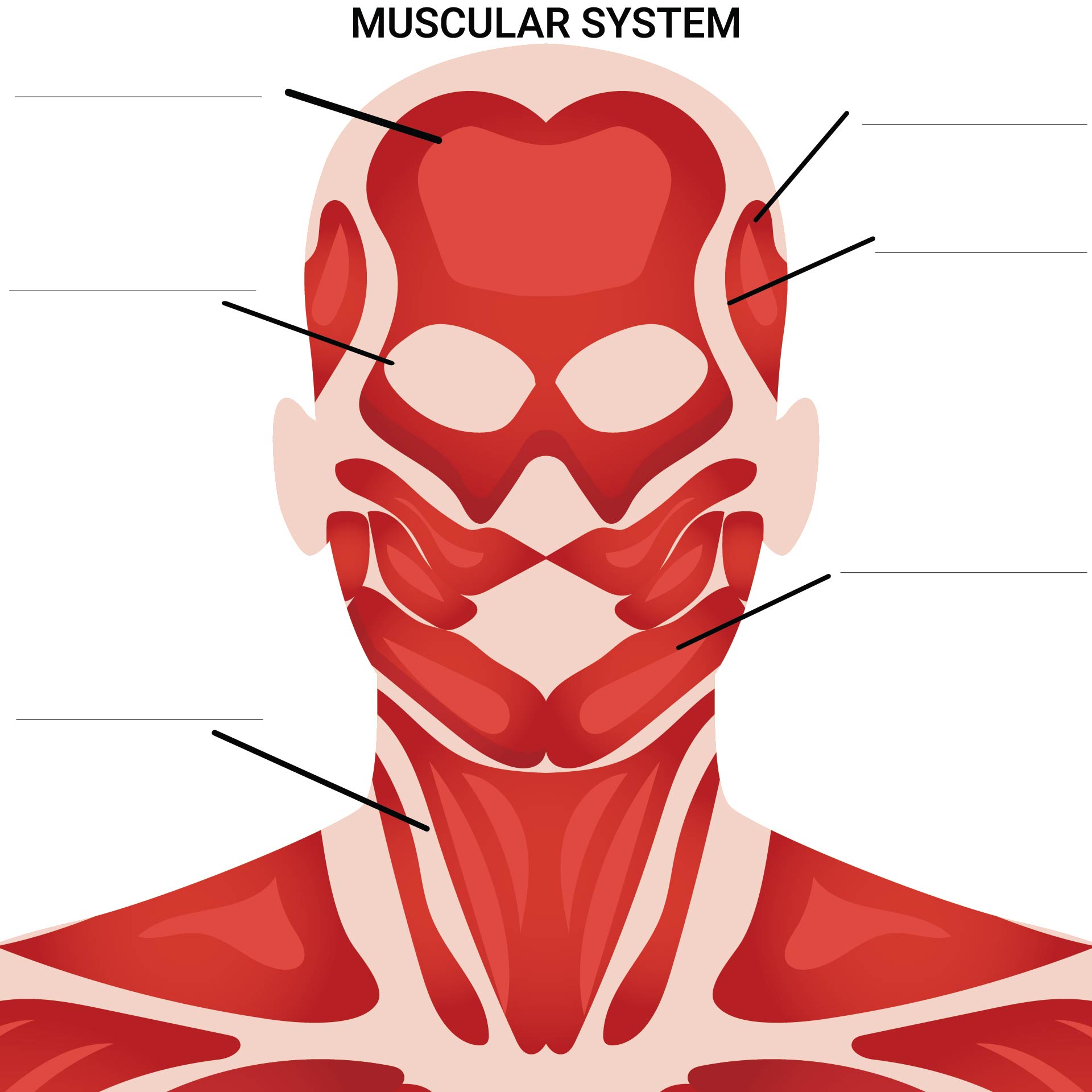
/ Blank head and neck muscles diagram muscular system diagram worksheet label muscles worksheet skull bones unlabeled anatomy. . Front and back muscles of the body. Everyone should list the structures within muscle. Now label the diagram in your workbook! The muscles extend from the tubercles of the ribs behind, to the cartilages of the ribs in ...
Dec 14, 2021 · Muscles of the neck (Musculi cervicales) The muscles of the neck are muscles that cover ... Definition And Function. A Large Group Of Muscles In Th ... Anterior muscles of the neck. Superficial muscles: Platysma, ... Lateral (vertebral) muscles of ... Scalene muscles: Anterior scal ...
Anatomy, Head and Neck, Buccinator Muscle; Free Review Questions. Introduction. The Buccinator muscle is a bilateral square-shaped muscle constituting the mobile as well as the adaptable cheek area. Couper and Myot coined the term buccinator in the year 1694.
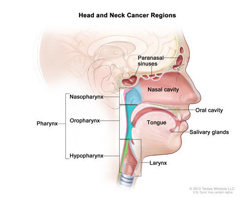
This chapter gives an overview of the important structures, muscles, fasciae. , and vessels ( arteries, veins, lymph. , nerves) of the head and neck region. The brain, one of the most important organs, is protected by the skull, both of which are covered in other articles. There are also individual articles for the organs of perception as well ...
This book is part of a series that is designed to help medical student to. study via solving questions, to get the maximum benefit, one shall study from a. rich and knowledgeable textbook first ...
The muscles of the neck can be divided into 3 groups: anterior, lateral, and posterior neck muscles. Each of the groups is subdivided according to function and the precise location of the muscles. The muscles of the neck are mainly responsible for the movements of the head (i.e., extension, flexion, lateral flexion-extension, and rotation), but ...
Muscles of the Head and Neck. Humans have well-developed muscles in the face that permit a large variety of facial expressions. Because the muscles are used to show surprise, disgust, anger, fear, and other emotions, they are an important means of nonverbal communication. Muscles of facial expression include frontalis, orbicularis oris, laris ...
Important Exam Questions on Head & Neck. To know the Answers, please click on the text highlighted in blue. Contents [ show] 1 Enumerate: 2 Draw labelled diagram to show: 3 Write short notes on: 4 Long questions: 5 Give anatomical basis of: 6 Share this:
Feb 22, 2012 - This Pin was discovered by Morgan Raimondo. Discover (and save!) your own Pins on Pinterest
Head and Neck mcqs 1) Regarding the superior orbital fissure, which is INCORRECT? a) its common tendinous ring binds the SOF content of nerves and muscles to the contents of the optic canal b) the origin of levator palpebrae superioris is its bony upper margin c) lacrimal, frontal and trochlear nerves pass through it ...
> Head and Neck Quiz 1 May 14, 2018 Anatomy , Head and Neck external carotid artery , external jugular vein , internal jugular vien , MCQs on head and neck , Muscles of mastication , nerve supply of tongue , parotid gland , scap dangerous layer POONAM KHARB JANGHU
The muscles of mastication are a group of muscles responsible for chewing (i.e. movement of the mandible at the temporomandibular joint). These muscles originate from the surface of the skull and insert onto the mandible.¹. There are four muscles that comprise the muscles of mastication, including masseter, temporalis, lateral pterygoid and medial pterygoid.¹
Neck muscles work together with tendons and ligaments to support and move the neck and head. Tendons are connective tissue that attach muscle to bone, whereas ligaments attach bones to other bones.
Blank head and neck muscles diagram lower leg muscle diagram blank and anatomy muscle coloring worksheet are some main things we want to present to you based on the post title. Free Anatomy Quiz The Muscular System Section. Start studying anatomy and physiology ch 1 body regions worksheet. Blank Muscle Diagram Worksheet.
A forward head posture can be the result of injuries like sprains and strains of the neck. It can also be created by: Weak neck muscles. Poor sleeping positions. Improper breathing habits. Rotational athletics where one side of the body is dominantly used (tennis, golf, hockey and baseball) Repetitive: Computer and TV use.
The omotransversarius muscle of the cow locates at the lateral surface of the neck extending from the wing of the atlas to the shoulder. You will find a thin brachiocephalic muscle that extends along the side of the neck from the head to the arm of a cow. The division of pectoralis superficialis muscles is not so distinct as in horses.
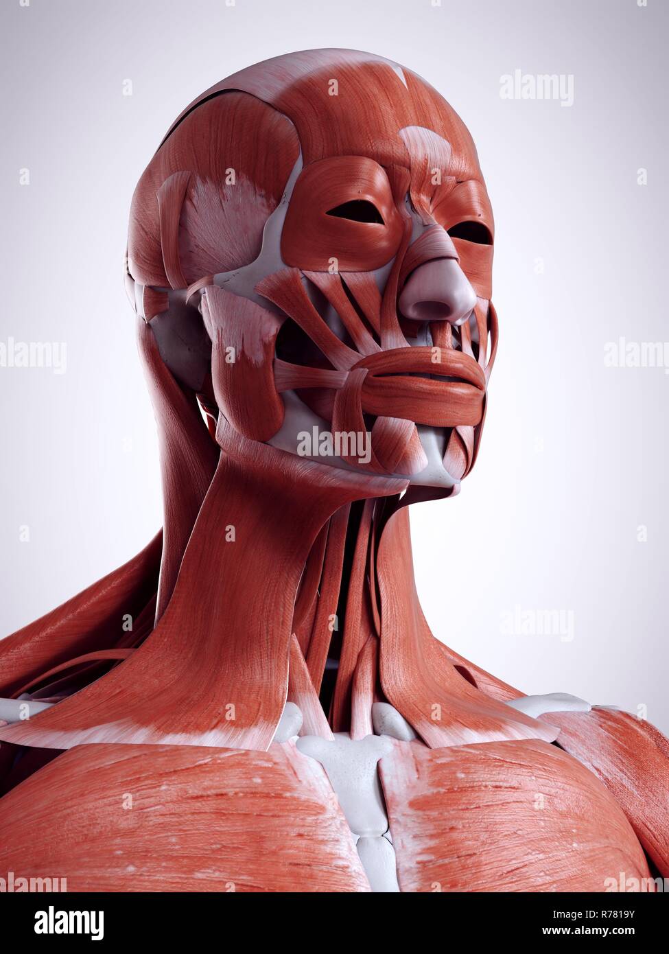
April 24, 2020 - Superficial muscles are the muscles closest to the skin surface and can usually be seen while a body is performing actions. Many in the neck help to stabilize or move the head. Some also create facial expressions.
The suboccipital muscles act to rotate the head and extend the neck. Rectus capitis posterior major and Rectus capitis posterior minor attach the inferior nuchal line of the occiput to the C2 and C1 vertebrae respectively. Obliquus capitis superior also extends from the occiput to C1 while ...
Instant anatomy is a specialised web site for you to learn all about human anatomy of the body with diagrams, podcasts and revision questions
Start studying Muscles of the Head and Neck - Head Model. Learn vocabulary, terms, and more with flashcards, games, and other study tools.
Neck neck muscle anatomy muscle diagram inspirational medical. To know the answers, please click on the text highlighted in blue. The splenius capitis and splenius cervicis, which are located in the back of the neck, work to rotate the head. Rotation and lateral flexion of the head are accomplished by lateral and posterior neck muscles.
MusFIG 5A & B: Muscle arms of the head muscles. A. Diagram showing two stylized head muscle arms (dark green) approaching nerve ring. Muscle arms from the 32 muscles in the head and neck project onto the inside surface of the nerve ring in a highly ordered fashion.
The trapezius muscle is a large muscle bundle that extends from the back of your head and neck to your shoulder. It is composed of three parts: The trapezius, commonly referred to as the traps, are responsible for pulling your shoulders up, as in shrugging, and pulling your shoulders back during scapular retraction.
27/06/2009 · Blank Head and Neck Muscles Diagram. Why is it important to learn muscle anatomy? Muscle and anatomy are two words that are often heard when you are studying science. The human body consists of many muscles. If someone wants a healthy and good life, one must understand his body. How do you take care of a body if you don't know the anatomy? …
There are six primary regions of lymph nodes - head and neck, axillary, upper limb, iliac, inguinal, and lower limb. Superficial and deep nodes run along the base of the head and throughout the neck.
January 21, 2018 - Neck muscles are bodies of tissue that produce motion in the neck when stimulated. The muscles of the neck run from the base of the skull to the upper back and work together to bend the head and assist in breathing.
Human body muscle diagram worksheet label muscles worksheet and blank head and neck muscles diagram are three of main things we want to present to you based on the post title. By the way about Muscle Labeling Worksheets Answers and Blanks we already collected several similar photos to add more info. Some of the worksheets displayed are Students ...
related to: neck muscles anatomy. betahealthy.com. 15 Common Causes of Neck Pain - Warning Signs & Early Symptoms. betahealthy.com has been visited by 100K+ users in the past month . Neck pain is something that is highly likely to be experienced by everyone at multiple. If, however, you end up experiencing severe pain, or the pain is lasting ...
Various head and neck muscles associated with chronic migraine: Botox is indicated for the treatment of chronic migraine, with injection sites varying based on patient-specific presentations and pain sites. Some migraine treatments may require injections to the muscles of the forehead, side of the head, and neck, while others may be limited to ...
splenius capitis. broad superficial muscle extending from upper thoracic vertebrae to skull, known as bandage muscle because it covers and holds down deeper neck muscles, origin- spinous processes of vertebrae C7-T6, insertion- mastoid process of temporal bone and occipital bone, extends or hyperextends head. sternocleidomastoid.
Muscles of the neck (Musculi cervicales) The muscles of the neck are muscles that cover the area of the neck.These muscles are mainly responsible for the movement of the head in all directions. They consist of 3 main groups of muscles: anterior, lateral and posterior groups, based on their position in the neck.The musculature of the neck is further divided into more specific groups based on a ...
Muscles of the Head and Neck - Listed Alphabetically. Muscle, Origin, Insertion, Action, Innervation, Artery, Notes, Image.
Even the middle ear takes part in the muscular system of the head and neck. In fact, the smallest muscle of the skeleton is the stapedius, which measures around 1 millimeter (1/20th of an inch) in length. The muscles of the middle ear contract to dampen the amplitude of vibrations from the eardrum to the inner ear.
Cases. By sharing our collective experience through interesting patient cases, we can make a real difference in how people are imaged and diagnosed. Each case belongs to a contributing member, which can then be viewed and added to articles or playlists by the community, and is guided by dedicated editors to match quality standards and privacy ...
Feb 23, 2014 - Muscles of The Head - Learn Human Anatomy
Head and neck (anterior view) The head and neck are two examples of the perfect anatomical marriage between form and function, mixed with a dash of complexity. The neck is resilient enough to sustain a five kilogram weight 24/7, yet sufficiently mobile to move it in several directions.



:background_color(FFFFFF):format(jpeg)/images/library/10461/cover_head_neck.png)

:background_color(FFFFFF):format(jpeg)/images/library/12652/Introduction.png)








:background_color(FFFFFF):format(jpeg)/images/library/14150/Neck_muscles.png)
0 Response to "8 head and neck muscle diagram"
Post a Comment