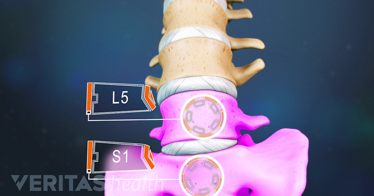
39 spine l5 s1 diagram
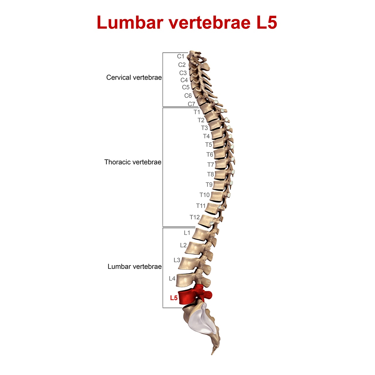
Human Spine and Spinal Cord Picture C1 - S5 Vertebra Information and pictures of the spine and spinal cord showing C1 to S5 vertebra and which The human spine is composed of 33 vertebrae that interlock with each other to form the spinal column. Fig 1. Lateral labeled diagram of the human vertebral spinal column showing vertebrae numbering... en.wikipedia.org › wiki › Thoracic_vertebraeThoracic vertebrae - Wikipedia In vertebrates, thoracic vertebrae compose the middle segment of the vertebral column, between the cervical vertebrae and the lumbar vertebrae. In humans, there are twelve thoracic vertebrae and they are intermediate in size between the cervical and lumbar vertebrae; they increase in size going towards the lumbar vertebrae, with the lower ones being a lot larger than the upper.
› itmat › eventsEvents | Institute for Translational Medicine and ... Symposia. ITMAT symposia enlist outstanding speakers from the US and abroad to address topics of direct relevance to translational science. Read more

Spine l5 s1 diagram
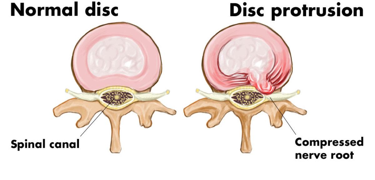
Treating an L5-S1 Disc Herniation: A Case Study - Regenexx Join us for a free Regenexx Spine Webinar. What Happens When an Epidural Doesn't Work? As I said above, his doctors were thrown for a loop When you have an L5-S1 disc bulge or herniation, that irritates the local L5 or S1 spinal nerves. Some parts of these nerves go down the leg causing sciatica. › scoliosis-curve-directionLevoscoliosis and Dextroscoliosis Scoliosis Directions Nov 25, 2020 · An X-ray is an important part of diagnosing scoliosis and determining the location and extent of spine misalignment. In the X-ray above, there is an area of dextroscoliosis and an area of levoscoliosis. In this image of an X-ray, the thoracic spine (top part) shows a dextroscoliosis, and the lumbar spine (bottom part) shows a levoscoliosis. en.wikipedia.org › wiki › Superior_cluneal_nervesSuperior cluneal nerves - Wikipedia Anatomy. The superior cluneal nerves are a group of nerves that were first described by Maigne et. Al in 1989 as a source of low back. These nerves are grouped as the superior cluneal nerves due to their trajectory over the iliac spine, as opposed to the lateral, medial and inferior cluneal nerves.
Spine l5 s1 diagram. L5-S1 Disc Degeneration - Causes and Treatments | Spinal stenosis Spine is also known as vertebral column, spinal column or backbone. In an adult, a vertebral column is constituted of 26 bones or vertebra. Having a brief idea of the structure of a spine, let us now discuss a lumbosacral joint. Diseases and Conditions Associated with L5-S1 region. All about L5-S1 (Lumbosacral Joint) | Spine-health The L5-S1 spinal motion segment helps transfer loads from the spine into the pelvis and legs. This motion segment receives a higher degree of mechanical L5 consists of a vertebral body in front and an arch in the back that has 3 bony protrusions: a prominent spinous process in the middle and two... Расположение протрузий C2-C7, Th1-Th12, L1-L5, L5-S1 » Клиника... Клиника Доктора Игнатьева » Протрузия » Расположение протрузий C2-C7, Th1-Th12, L1-L5, L5-S1. Добрий день.В мене на час обстеження МСКТ—ознаки остеохондрозу L1-S1 сегментів хребта.Задня дифузна протрузія міжхребцевого диску L4-L5..Задня центральна частково... en.wikipedia.org › wiki › Superior_cluneal_nervesSuperior cluneal nerves - Wikipedia Anatomy. The superior cluneal nerves are a group of nerves that were first described by Maigne et. Al in 1989 as a source of low back. These nerves are grouped as the superior cluneal nerves due to their trajectory over the iliac spine, as opposed to the lateral, medial and inferior cluneal nerves.
› scoliosis-curve-directionLevoscoliosis and Dextroscoliosis Scoliosis Directions Nov 25, 2020 · An X-ray is an important part of diagnosing scoliosis and determining the location and extent of spine misalignment. In the X-ray above, there is an area of dextroscoliosis and an area of levoscoliosis. In this image of an X-ray, the thoracic spine (top part) shows a dextroscoliosis, and the lumbar spine (bottom part) shows a levoscoliosis. Treating an L5-S1 Disc Herniation: A Case Study - Regenexx Join us for a free Regenexx Spine Webinar. What Happens When an Epidural Doesn't Work? As I said above, his doctors were thrown for a loop When you have an L5-S1 disc bulge or herniation, that irritates the local L5 or S1 spinal nerves. Some parts of these nerves go down the leg causing sciatica.




:background_color(FFFFFF):format(jpeg)/images/library/12522/spine-bones-and-ligaments-Recovered_english.jpg)


:max_bytes(150000):strip_icc()/discus-l5-s1-185295300-5bfd7badc9e77c0026f1b069.jpg)



0 Response to "39 spine l5 s1 diagram"
Post a Comment