42 long bone diagram labeled
- 2 blank long bone diagrams, one labeled, "Anatomy of a Long Bone", the other completely blank - 1 rough sketch of a bone slice, showing the structure of cancellous, (spongy), and compact bone, black and white - A simple, one page handout, with small black and white images, describing the different types of bone cells and what they do. - A 3 ... Jan 10, 2022 · Long Bone Diagram Labeled. Here are a number of highest rated Long Bone Diagram Labeled pictures on internet. We identified it from reliable source. Its submitted by management in the best field. We allow this kind of Long Bone Diagram Labeled graphic could possibly be the most trending subject bearing in mind we part it in google plus or facebook.
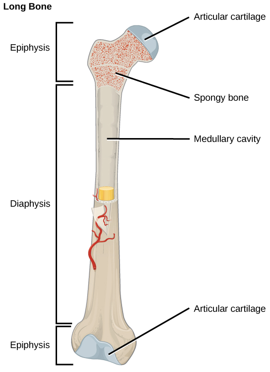
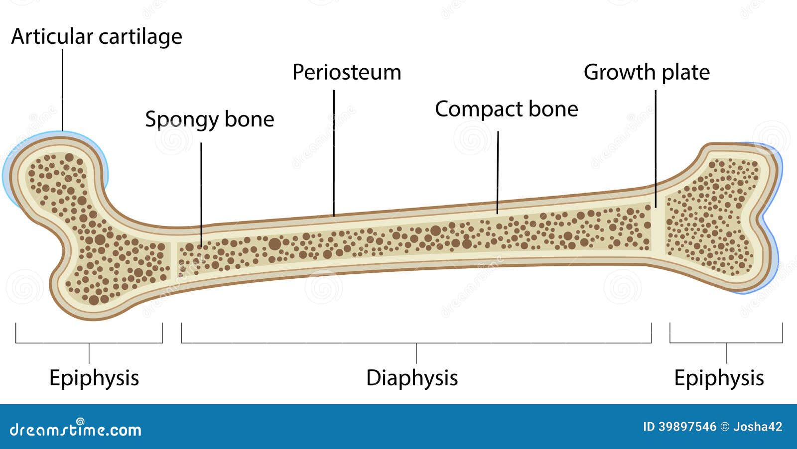
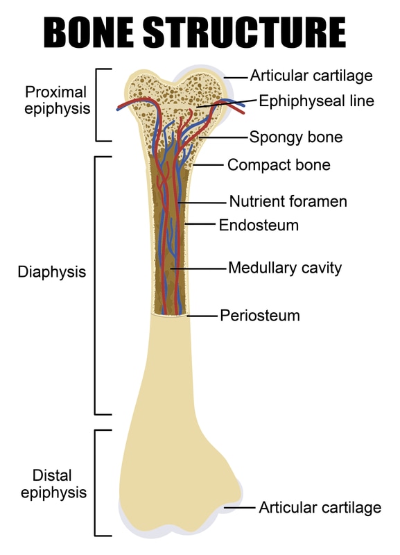
A long bone has two parts: the diaphysis and the epiphysis. The diaphysis is the tubular shaft that runs between the proximal and distal ends of the bone. The ...

Long bone diagram labeled
Humerus bone labeled vector illustration diagram Humerus bone labeled vector illustration diagram. Long bone type in the upper arm. Skeleton anatomy scheme with greater tubercle, deltoid tuberosity, medial epicondyle, trochlea and other parts. humerus labeled diagram stock illustrations. Male Biceps Muscles Isolated in Skeleton Labeled Chart on ... This is an online quiz called Label a Long Bone. There is a printable worksheet available for download here so you can take the quiz with pen and paper. Your Skills & Rank. Total Points. 0. Get started! Today's Rank--0. Today 's Points. One of us! Game Points. 13. You need to get 100% to score the 13 points available. A long bone is a bone that is significantly longer than it is wide. Examples of long bones are the femur, tibia, and fibula of the leg, the humerus, radius, and ulna of the arm, and the phalanges of the fingers and toes. A long bone consists of a long shaft (diaphysis) with two bulky ends or extremities where articulation takes place.
Long bone diagram labeled. The long bones are those that are longer than they are wide. They are one of five types of bones: long, short, flat, irregular and sesamoid. Long bones ... Long bone • Longer than they are wide. • Reflects the elongated shape rather than the overall size. • Consist of a shaft plus two ends and are constructed primarily of compact bone • may contain substantial amounts of spongy bone. • All bones of the limbs, except the patella, wrist and ankle bones, are long bones. 6. Femur Bone Anatomy. The femur is a type of long bone located in the thigh and is the largest bone of the skeletal system. There was a previous EZmed post (see below) on the anatomy of the femur where we labeled all of the main parts of the bone on a color-coded diagram. For the step-by-step video and blog post that walks through the anatomy of ... Apr 25, 2013 — A long bone has two parts: the diaphysis and the epiphysis. The diaphysis is the tubular shaft that runs between the proximal and distal ends of ...
Articular cartilage · dense, white, connective tissue that covers the articulating surfaces of bones at joint ; Epiphysis · the bulbous end of a long bone. The epiphyseal line is a remnant of an area that contained hyaline cartilage that grew during childhood to lengthen the bone. Long bones contain yellow bone marrow and red bone marrow, which produce blood cells. The thigh bone (femur) is a long bone. Some bones in the fingers are classified as long bones, even though they are short in length. Mar 29, 2021 · Femur Bone Anatomy. The femur is a type of long bone located in the thigh and is the largest bone of the skeletal system. The femur and/or hip may fracture secondary to trauma, so understanding the femur bone anatomy is important. The anatomy of the femur can be divided into proximal, central, distal, and posterior parts. The 5 main bone types in the human body skeletal system. Labeled diagrams and examples of long bones, short bones, flat bones, sesamoid bones, and irregular bones that make up the foot, hand, skull, cranium, arm, leg, ankle, wrist, hip, and vertebrae or spine.
30 seconds. Q. Periosteum. answer choices. the membrane lining the bone cavity. the tough membrane covering the shaft of the bone. the shiny, articulating cartilage on the ends of a bone. the blood vessels inside a bone. Tags: Question 8. The Long Bone Diagram Blank could be your desire when thinking of about Bone. When showing this Long Bone Diagram Blank, I can guarantee to rock your world!. For this time we collect some pictures of Long Bone Diagram Blank, and each of them giving you some fresh ideas. Femur Bone Parts With Diagram - See more about Femur Bone Parts With Diagram. Start studying Long bone labeled. Learn vocabulary, terms, and more with flashcards, games, and other study tools. Humerus bone labeled vector illustration diagram stock illustration. Save to Board. Humerus bone labeled vector illustration diagram Humerus bone labeled vector illustration diagram. Long bone type in the upper arm. Skeleton anatomy scheme with greater tubercle, deltoid tuberosity, medial epicondyle, trochlea and other parts. Humerus stock vector.
19.2 Bone - Concepts of Biology - 1st Canadian Edition
A long bone has two parts: the diaphysis and the epiphysis. The diaphysis is the tubular shaft that runs between the proximal and distal ends of the bone. The ...

Bones. Bones Structure. Bone Tissue. Bone Membranes
Oct 28, 2021 · Forearm muscles (extensors) labeled and unlabeled. To begin, spend some time looking at the forearm muscles diagram above. Here you can see all the extensor forearm muscles clearly labeled. Once you’re ready, you can try labeling the muscles for yourself using the blank forearm muscles diagram free to download below.

Long Bone Diagram Labeled / Long Bone Images Stock Photos ...
Elements [1-36] 20 terms. Kirulus_Fanous TEACHER. Long Bone Anatomy, Anatomical Terminology. 38 terms. Kirulus_Fanous TEACHER. Upgrade to remove ads. Only $2.99/month.
What is the structure of a long bone - L2 and L3 anatomy ...
Gross Anatomy of Bones. A long bone has two main regions: the diaphysis and the epiphysis ( Figure 6.3.1). The diaphysis is the hollow, tubular shaft that runs between the proximal and distal ends of the bone. Inside the diaphysis is the medullary cavity, which is filled with yellow bone marrow in an adult.

Basic Long Bone Diagram Labeled : Structure and Function ...
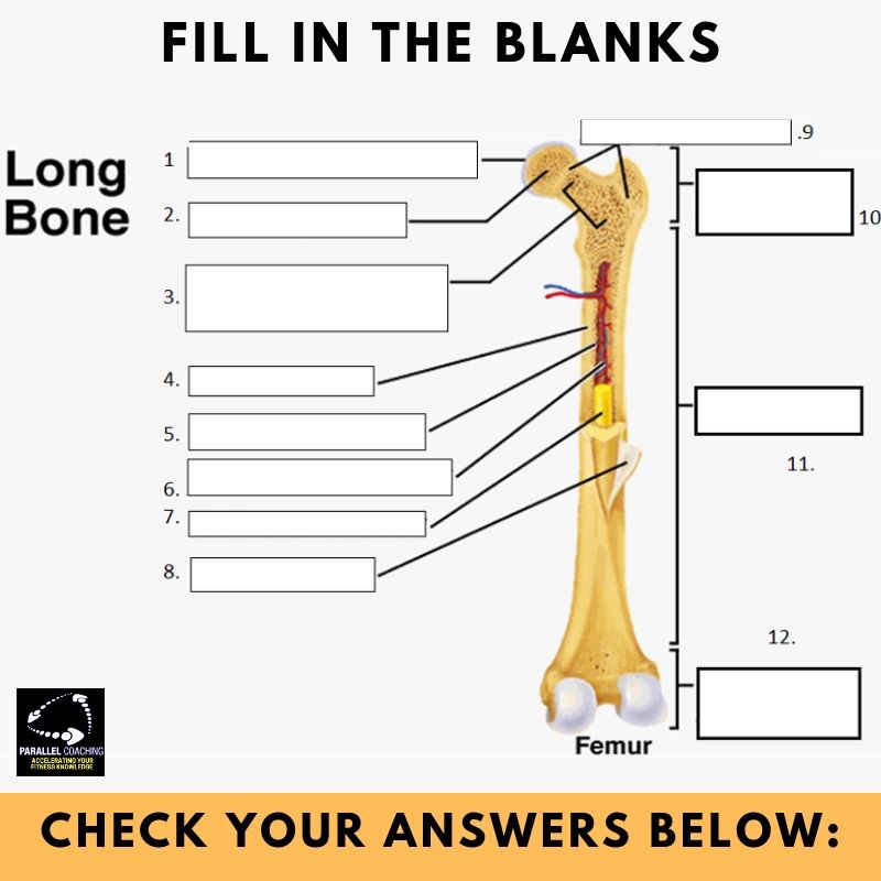
Anatomy of a Long Bone. Instructions: Read the text. Use the text and the diagram to help you to answer the questions. The ends of long bones are called . epiphyses. Each epiphysis is shaped differently; each end is specially. designed to fit the bone or bones it attaches to. In the . diagram of the femur (thigh bone) on the right, notice

long bone diagram | timothyakeller | Flickr
Start studying Long Bone Anatomy. Learn vocabulary, terms, and more with flashcards, games, and other study tools.

Image from page 184 of "The American Museum journal" (c1900-[1918])
Start studying LABEL A LONG BONE (HUMERUS). Learn vocabulary, terms, and more with flashcards, games, and other study tools.

Gross anatomy of the typical long bone in 2020 | Gross ...
Start studying Anatomy of a Long Bone. Learn vocabulary, terms, and more with flashcards, games, and other study tools.
Structure of bone | Science online
Find Long bone anatomy stock images in HD and millions of other royalty-free stock photos, illustrations and ... Labeled anatomical skeleton set scheme.

Pair of jellyfish
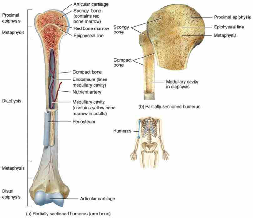
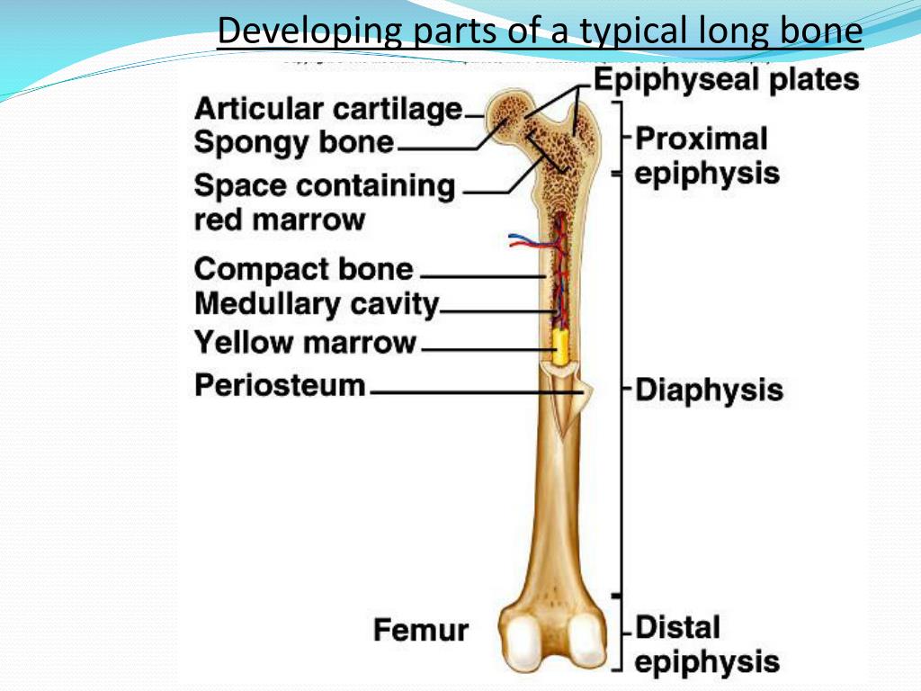
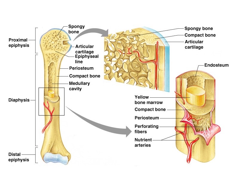
Oct 01, 2019 · Parts of long bones. This image represents the parts of a long bone. The labels include proximal epiphysis, proximal metaphysis, diaphysis (bone shaft), distal metaphysis, distal epiphysis, and epiphyseal line (x2). Structure of an adult human long bone. The following image gets into a little more detail in regard to human long bone structure.
Anatomy Of A Typical Long Bone | MedicineBTG.com
Long bone model compact bone diagram labeled anatomy human body picture of long bone labeled long bone diagram anatomy organ. The long bones are those that are longer than they are wide. Examples of long bones are the femur tibia and fibulaof the leg the humerus radius and ulnaof the arm and the phalangesof the fingers and toes.

Black Rabbit, Byker Farm, Ouseburn Valley, Newcastle Upon Tyne, Tyne & Wear, England.
Anatomy of Long Bone. Share Share by Harrisonk102. G10 G11 G12 Anatomy. Like. Edit Content. Embed. More. Log in required. Theme. Fonts: Log in required. Options. Leaderboard. Show more Show less . This leaderboard is currently private. Click Share to make it public. This leaderboard has been disabled by the resource owner. ...

Long Bone Labeled Diagram - Bone Structure Anatomy And ...
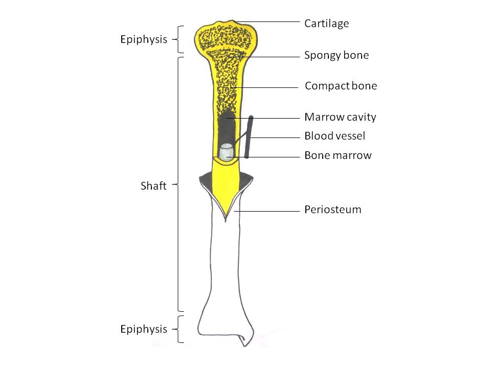
Anatomy of Long Bones The long bones have a long, central shaft that enlarges at the ends into epiphysis.The long bones in the legs are the femur, tibia, and fibula. The long bones of the arms are the radius and ulna. Both the feet and hands have long bones in the digits – the phalanges.The adult femur is the largest bone in

Long Bone Labeled : 1 Schematic Drawing Of A Longitudinal ...
Long bones grow more than the other classes of bone throughout childhood and so are responsible for the bulk of our height as adults. A hollow medullary cavity is found in the center of long bones and serves as a storage area for bone marrow. Examples of long bones include the femur, tibia, fibula, metatarsals, and phalanges. Short. Short bones ...
Bone Anatomy Labeled Diagram Stock Vector - Illustration ...
The ilium is the big bone of the hip, the ischium is the bone on which one sits and the pubis forms the lower frontal hip bone as seen in the diagram. Femur. The longest and the strongest bone in the human skeletal system as you can observe in the labeled skeleton diagram of the human body. The femur or the thigh bone is closest to the body.

Jellyfish in the tank
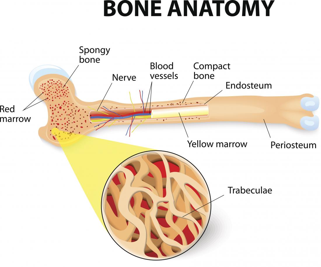
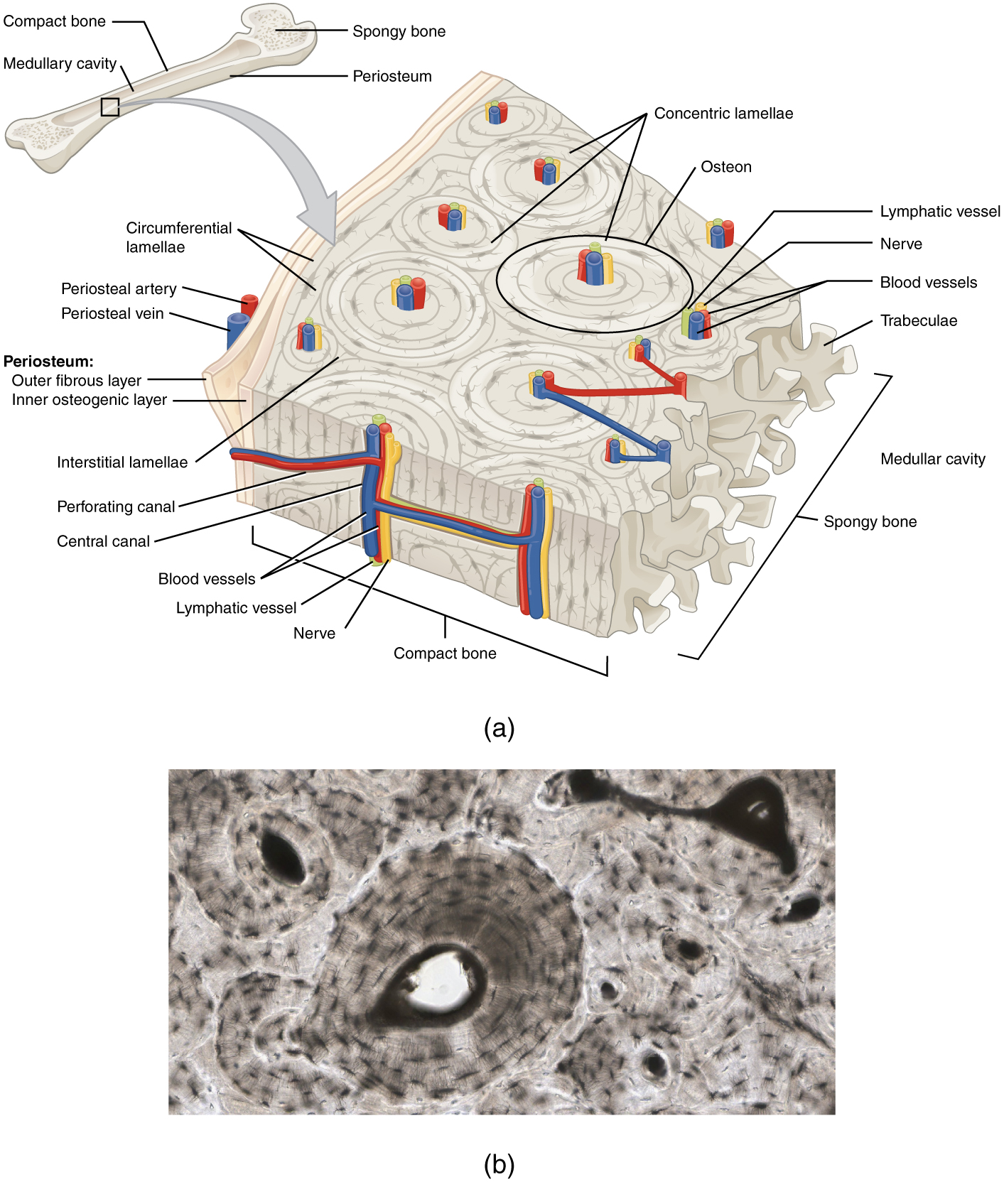
Parts of long bone (applies to other bones too). Used figure 6.2 in book. Some descriptions for confusing parts.Omit number 13 in the picture.

Long Bone Labeled : Long Bone Labeled Diagram Quizlet ...
Bone Diagram Forehead (Frontal bone) Nose bones (Nasals) Cheek bone (Zygoma) Upper jaw (Maxilla) Lower jaw (Mandible) Breast bone (Sternum) Upper arm bone (Humerus) Lower arm bone (Ulna) Thigh bone (Femur) Collar bone (Clavicle) Toe bones (Phalanges) Ankle bones (Tarsals) Kneecap (Patella) Shin bone

Long Bone Labeled Diagram Quizlet - Long Bone Diagram ...
A long bone is a bone that is significantly longer than it is wide. Examples of long bones are the femur, tibia, and fibula of the leg, the humerus, radius, and ulna of the arm, and the phalanges of the fingers and toes. A long bone consists of a long shaft (diaphysis) with two bulky ends or extremities where articulation takes place.

Long Bone Diagram Labeled Quizlet / Anatomy Lecture 5 ...
This is an online quiz called Label a Long Bone. There is a printable worksheet available for download here so you can take the quiz with pen and paper. Your Skills & Rank. Total Points. 0. Get started! Today's Rank--0. Today 's Points. One of us! Game Points. 13. You need to get 100% to score the 13 points available.
Microscopic Structure of Bone | ClipArt ETC
Humerus bone labeled vector illustration diagram Humerus bone labeled vector illustration diagram. Long bone type in the upper arm. Skeleton anatomy scheme with greater tubercle, deltoid tuberosity, medial epicondyle, trochlea and other parts. humerus labeled diagram stock illustrations. Male Biceps Muscles Isolated in Skeleton Labeled Chart on ...

Catoptromancy....(or Enoptromancy)..Looking into water's mirror to see the future or contact a supernatural entity...suspended by a thread till its based touched the surface of the water, having first prayed to the goddess and offered incense.

Long Bone Labeled Quizlet : Anatomy Bones Quizlet ...
34 Label The Parts Of A Long Bone - Labels Design Ideas 2020
31 Label The Parts Of A Long Bone - Label Ideas 2020
Chapter 6 Bones and Cartilage - Biology 4 Human ...

Light the way

Long Bone Diagram Compact Bone : structures of a long bone ...

Label the diagram of a long bone Quiz

Long Bone Labeled Epiphysis : Solved: Correctly Label The ...

Labeling Long Bone Structure / Introduction to Bone ...

Long Bone Diagram With Labels : Structure Of A Long Bone ...

Long Bone Labeled : Long Bone Labeled Anatomy File Human ...

Black Beauty, Equine, Harlow Green, Gateshead, Tyne & Wear, England.

Long Bone Labeling - Long Bone Label The Structure The ...

Bitch, Long Beach, Iskele, Turkish Republic Of North Cyprus.
Structure and functions of bones - Online Science Notes

Testosterone...A testicular action was linked to circulating blood fractions – now understood to be a family of androgenic hormones – in the early work on castration and testicular transplantation in fowl

Basic Long Bone Diagram Labeled : Structure and Function ...

Structure of Compact Bone Quiz

Humerus - Medical Art Library

Figure 11.8. Anatomy of a Long Bone (Humerus)
A generic long bone is shown at the top of this ...
0 Response to "42 long bone diagram labeled"
Post a Comment