40 compound microscope ray diagram
AN INTRODUCTION TO THE COMPOUND MICROSCOPE OBJECTIVE: In this lab you will learn the basic skills needed to stain and mount wet slides. You will also learn about magnification, resolution and the parts of the compound microscope. INTRODUCTION: The light microscope can extend our ability to see detail by 1000 times, so that we can Transmission electron microscopy (TEM) analysis is conducted to get the actual size of the nanocrystalline cellulose fibers and in some cases the morphology. Nasseri and Mohammadi [99] obtained individual cellulose whiskers with length (L) of 87±28 nm and diameter (d) of 15±3 nm, with an average aspect ratio (L/d) of whiskers obtained was 6±2.. Jiang and Hsieh obtained cellulose NFs …
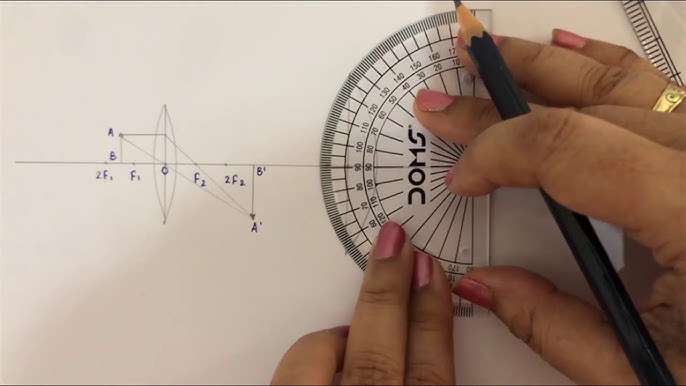
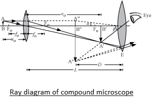
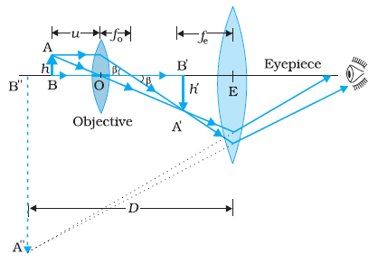
(a) Draw a ray diagram for the formation of image by a compound microscope. (b) You are given the following three lenses. Which two lenses will you use as an eyepiece and as an objective to construct a compound microscope? Lenses Power (D) Aperture (cm) L1 3 8 L2 6 1 L3 10 1 (c) Define resolving power of a microscope and write one factor on which it depends.
Compound microscope ray diagram
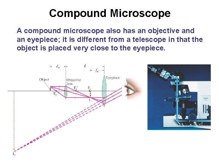
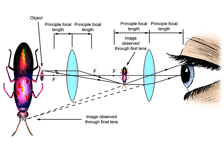
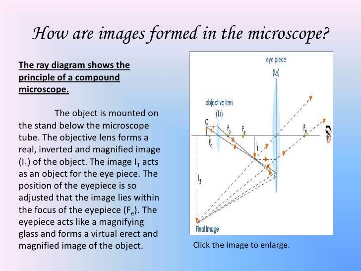
A highly magnified image is thus formed. Complete step-by-step answer: A compound microscope consists of two converging lens systems, there is an objective lens ...1 answer · Top answer: Hint: A compound microscope is an optical instrument used for observing highly magnified images of tiny objects. The compound microscope has two lenses ... (b) Show that in order to achieve large magnification in a compound microscope the magnitude of focal length of objective and eye piece should be small. Medium.1 answer · Top answer: (A) : Compound microscope is consist of two convex lenses, one object of very small focal length with short aperture and one eyepiece, E of moderate ... Here are two ray diagrams for compound microscope, the first one proposed by the book, and the second one recommended by the teacher: View attachment 232683 View attachment 232684 In the first image, the light rays form a real image A'B', which becomes the virtual object for the eyepiece. See, the original rays are carried forward to the ...
Compound microscope ray diagram. A ray diagram from left to right shows a virtual inverted enlarged final image of the. Figure 2. A compound microscope composed of two lenses, an objective ... 18.04.2021 · A simple or compound microscope is made of two important parts such as the Structural parts and Optical parts. The structural parts provide supports to the microscope, while the optical parts help to produce a magnified image of specimens. The structural parts of Microscope with their functions. This portion of microscope is made of three important parts such as Head, base and arm. Each of ... A Draw A Ray Diagram Showing The Image Formation By A Compound. Magnification Of Compound Microscope With Derivation And Diagram. Solved The Focal Length Of A Compound Microscope S Object. Notes On Microscope Grade 11 Physics Optical Instruments. The Compound Microscope Physics Homework Help Physics Assignments. Compound microscope is a type of optical microscope that is used for obtaining a high-resolution image. There are more than two lenses in a compound microscope. Learn about the working principle, parts and uses of a compound microscope along with a labeled diagram here.
7 Dec 2020 — draw a labelled ray diagram showing imagr formation in compound microscope.define its magnifying power and write expression for it. Geometrical Construction of Ray Diagrams. A popular method of representing a train of propagating light waves involves the application of geometrical optics to determine the size and location of images formed by a lens or multi-lens system. This tutorial explores how two representative light rays can establish the parameters of an imaging scenario. Draw a neat labeled ray diagram of a compound microscope. Explain briefly its working. asked Aug 1, 2019 in Physics by Rk Roy (63.7k points) jee; jee mains; 0 votes. 1 answer. Draw a neat labelled diagram of a compound microscope and explain its working. asked Nov 21, 2019 in Physics by Raghab (50.5k points) class-11; ADVERTISEMENTS: Read this article to learn about the working principle and parts of a compound microscope with diagrams! Working Principle: The most commonly ...
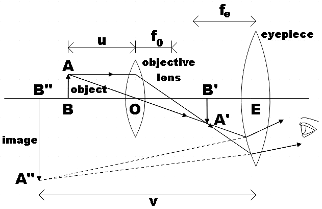
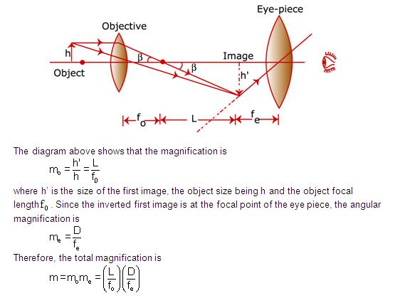
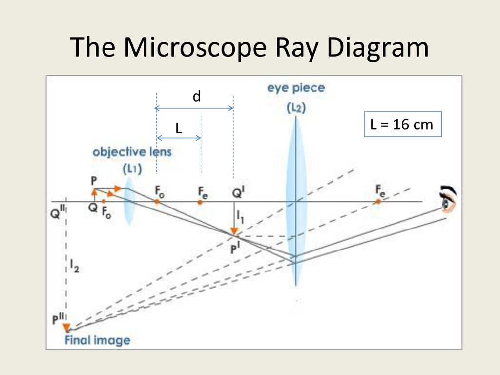
Here is the ray diagram of a compound microscope. So, when we are focussing, we move the objective lens which tweaks the image distance. My doubt is that, shouldn't the image be seen clearly, wheresoever the first real image forms, if within Fe (Focus of the eyepiece lens). distance or image size. Thus, a ray diagram is used to find the final image distance and to determine the total magnification. The overall magnification of a compound microscope is the product of the individual magnifications of each lens: m = mome (1) where the magnification of either lens is given by: m = - q/p (2) Draw a ray diagram of a compound microscope for the final image formed at least distance of distinct vision.1 answer · Top answer: (a) A microscope is a device used to see magnified image of very small things which, a compound microscope consists of two convex lenses namely eyepiece ... The optical microscope, also referred to as a light microscope, is a type of microscope that commonly uses visible light and a system of lenses to generate magnified images of small objects. Optical microscopes are the oldest design of microscope and were possibly invented in their present compound form in the 17th century. Basic optical microscopes can be very simple, although many …
How To Draw Perfect Compound Microscope Ray Diagram In Minutes Board Exam Short Trick Std10 12 Youtube
Transmission electron microscopy (TEM) is a microscopy technique in which a beam of electrons is transmitted through a specimen to form an image. The specimen is most often an ultrathin section less than 100 nm thick or a suspension on a grid. An image is formed from the interaction of the electrons with the sample as the beam is transmitted through the specimen.
The Final Image Formed By A Compound Microscope Is Class 12 Physics Cbse
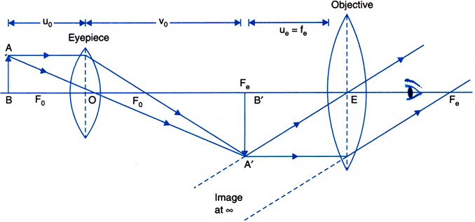
Illustrated in Figure 13 is the optical train, using ray traces, of an infinity-corrected microscope system. The components of this system are labeled in a similar manner to the finite-tube length system (Figure 12) for easy comparison. Here, the magnification of the objective is determined by the focal length of the tube lens. Note the infinity "afocal" space that is defined by parallel light ...
Phys 102 Lecture 2324 Lenses And Optical Instruments
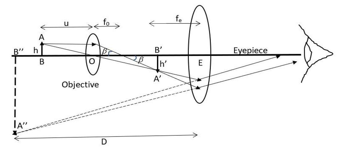
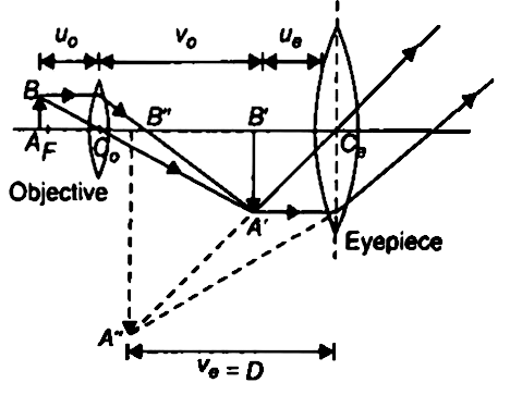
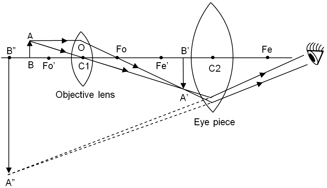
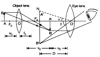
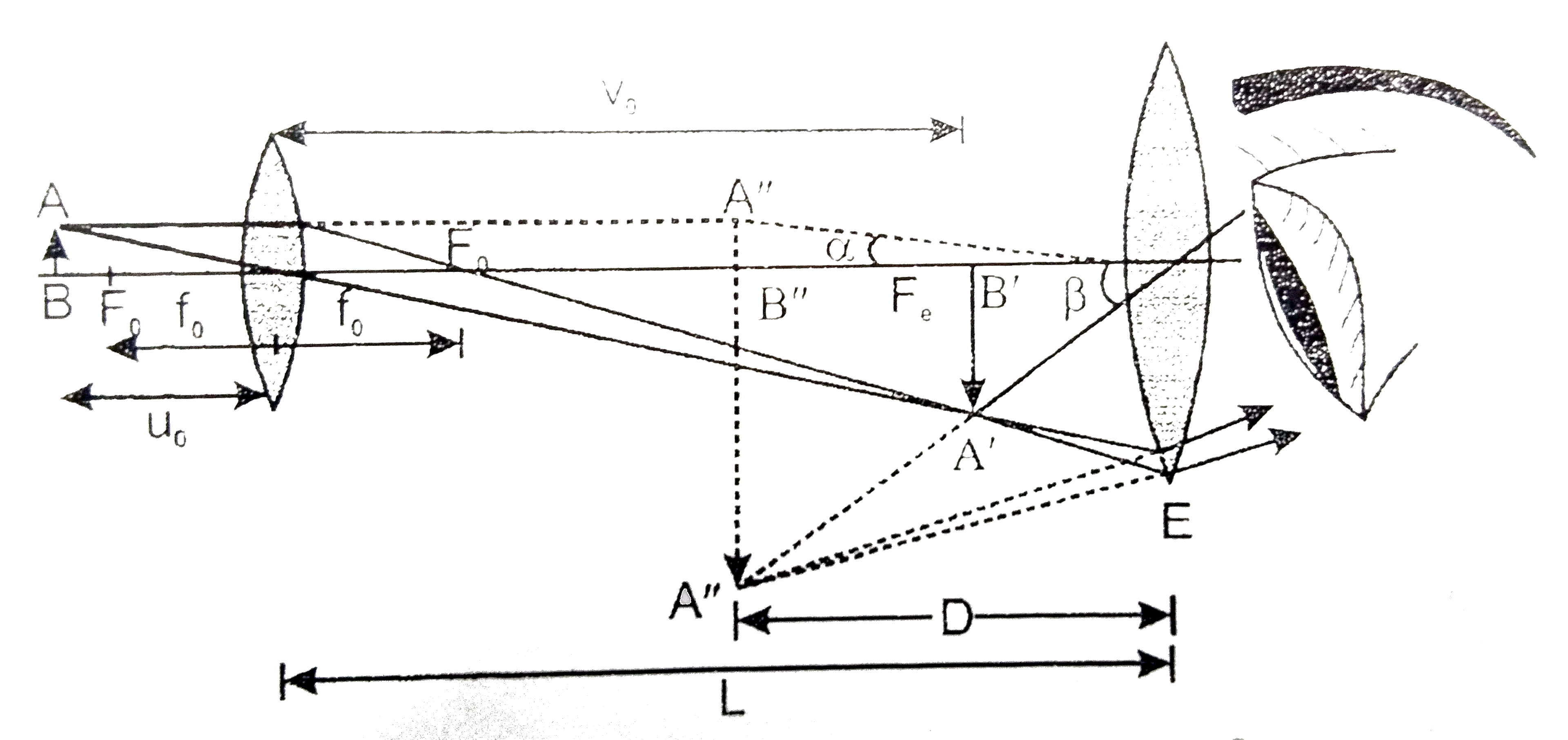
Ray diagram of a compound microscope.When the final image is formed at the least distance of distinct vision,For the image formed at infinity, ue = feand By making focal length of the objective small, the magnifying power can be increased.

Cutul With The Help Of A Ray Diagram Derive An Expression For Magnifying Power Of A Compound Microscope When The Image Is Formed At Near Point In A Compound Microscope An Object
21.10.2021 · The phase compositions of the compound coatings are shown in Fig. 2.Three intermetallic compounds were formed at 950–1200 °C, identified as HfIr 3, HfIr, and Hf 2 Ir, respectively. This result is consistent with the phase diagram of the Ir Hf binary alloys. Five diffraction peaks can be observed at 2θ = 40.66, 47.30, 69.13, 83.41, and 88.04°, corresponding to the (111), (200), (220), (311 ...

Experts Please Give Ray Diagram Of Compound Microscope Physics Ray Optics And Optical Instruments 14283469 Meritnation Com
Here are two ray diagrams for compound microscope, the first one proposed by the book, and the second one recommended by the teacher: View attachment 232683 View attachment 232684 In the first image, the light rays form a real image A'B', which becomes the virtual object for the eyepiece. See, the original rays are carried forward to the ...

A Draw A Ray Diagram Of Compound Microscope For The Final Image Formed At Least Distance Of Distinct Vision B An Physics Ray Optics And Optical Instruments 11967274 Meritnation Com
(b) Show that in order to achieve large magnification in a compound microscope the magnitude of focal length of objective and eye piece should be small. Medium.1 answer · Top answer: (A) : Compound microscope is consist of two convex lenses, one object of very small focal length with short aperture and one eyepiece, E of moderate ...

I Draw A Schematie Ray Diagram Ot A Compound Microscope When Image Is Formed At Distance Of Distinct Vision Ii Write The Expression For Resolving Power Of A Compound Microscope How Can
A highly magnified image is thus formed. Complete step-by-step answer: A compound microscope consists of two converging lens systems, there is an objective lens ...1 answer · Top answer: Hint: A compound microscope is an optical instrument used for observing highly magnified images of tiny objects. The compound microscope has two lenses ...
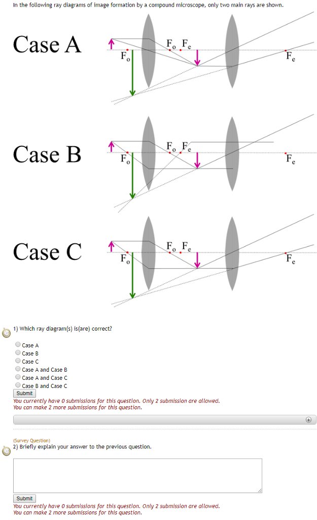
Solved In The Following Ray Diagrams Of Image Formation By A Chegg Com

A Draw A Labelled Ray Diagram Showing The Formation Of A Final Image By A Compound Microscope Youtube
Compound Microscope Ray Diagram Mistakes Physics Forums
Draw A Ray Diagram Of Compound Microscope When The Class 12 Physics Cbse
Draw A Ray Diagram Of A Compound Microscope Write The Expression For Its Magnifying Power From Physics Ray Optics And Optical Instruments Class 12 Cbse

Ray Diagram Of A Compound Microscope In 2021 Diagram Microscope Principles
Draw A Ray Diagram Showing Image Formation In A Compound Microscope Define The Term Limit Of Resolution And Name The Factors On Which It Depends How Is It Related To Resolving Power

Draw A Ray Diagram Of A Compound Microscope Diagram Transparent Png 678x317 Free Download On Nicepng

A Draw A Ray Diagram For The Formation Of Image By A Compound Microscope B You Are Given The Following Three Lenses Which Two Lenses Will You Use As An Eyepiece And
Draw A Labelled Ray Diagram Of A Compound Microscope And Explain Its Working Home Work Help Learn Cbse Forum
Compound Microscope Ray Diagram Mistakes Physics Forums
Which Ray Diagram Is Correct For A Compound Microscope Physics Forums
Derive The Formula For Angular Magnification Of A Compound Microscope When The Final Image Is Formed At Least Distance Of Distinct Vision Draw The Required Ray Diagram From Physics Microscopes And Telescopes
Compound Microscope Ray Diagram Mistakes Physics Forums

Or Draw Ray Diagram Of A Compound Microscope Showing The Final Image Formed At The Least Distance Of The Distinct Vision And Derive The Formula Of The Magnifying Power Of This Position

Draw A Ray Diagram To Show Formation Of An Image By A Compound Microscope Or An Astronomical Telescope Brainly In
Explain The Construction And Working Of A Compound Class 12 Physics Cbse
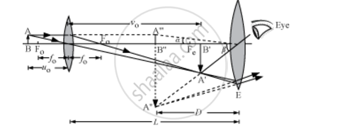
Draw A Labelled Ray Diagram Showing The Formation Of A Final Image By A Compound Microscope At Least Distance Of Distinct Vision Physics Theory Shaalaa Com
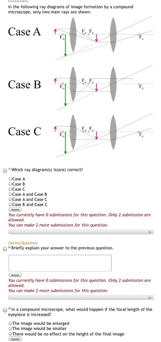
Solved In The Following Ray Diagrams Of Image Formation By A Chegg Com
1

How To Draw Compound Microscope Diagram Final Image At D Youtube

Draw A Neat Labelled Diagram Of A Compound Microscope And Explain Its Working Derive An Expression For Its Magnification
A Draw A Ray Diagram Showing The Image Formation By A Compound Microscope Hence Obtain Expression For Total Magnification When The Image Is Formed At Physics Topperlearning Com 7y05qdrr

A Draw A Labelled Ray Diagram Of Compound Microscope When Final Image Forms At The Least Distance Of Distinct Vision B Why Is Its Objective Of Short Focal Length And Of Short
Convex Lens Use Microscope

Leed Lens And Ray Diagram Electron Microscope Diagram Transparent Png 592x717 Free Download On Nicepng
The Microscope
Draw A Ray Diagram To Show The Working Of A Compound Microscope Deduce An Expression For The Total Magnification The Final Image Is Formed At The Near Point In A Compound Microscope

Kopal Classes Compound Microscope Ray Diagram Facebook

Draw A Labelled Ray Diagram Of An Image Formed By Compound Microscope When The Final Image Lies At The Least Distance Of Distinct Vision D

Draw The Ray Diagram Of Image Formation In Case Of Compound Microscope
Draw A Labelled Ray Diagram Showing Imagr Formation In Compound Microscopedefine Its Magnifying Power And Write Expression For It Physics Topperlearning Com Rn7zxv55
The Compound Microscope Ppt Download
0 Response to "40 compound microscope ray diagram"
Post a Comment