38 cardiac muscle tissue diagram
We'll also cover the benefits of exercise for cardiac muscle tissue. ... Use this interactive 3-D diagram to explore the movement of cardiac muscle tissue.Images · Cardiomyopathy · Exercise · Takeaway This diagram illustrates the molecular mechanism of muscular contraction. With application of a stimulus,. Muscle Contraction and Actin-Myosin Interactions: ...
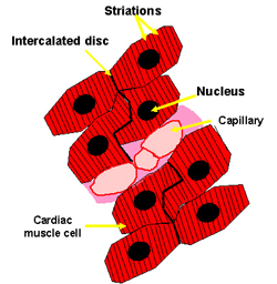
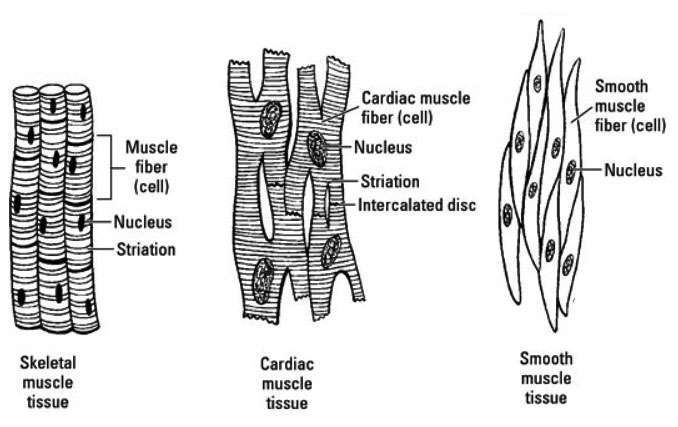
Cardiac Muscle Tissue. Cardiac muscle tissue is found only in the heart, where cardiac contractions pump blood throughout the body and maintain blood pressure. As with skeletal muscle, cardiac muscle is striated; however it is not consciously controlled and so is classified as involuntary. Cardiac muscle can be further differentiated from skeletal muscle by the presence of intercalated discs ...

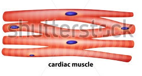
Cardiac muscle tissue diagram
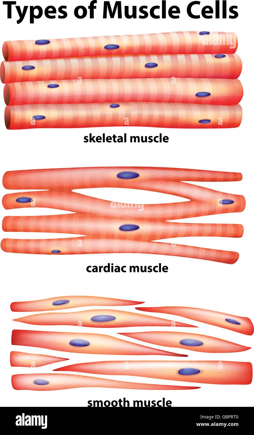
Cardiac muscle cells usually have a single (central) nucleus. The cells are often branched, and are tightly connected by specialised junctions. The region where ... 28.10.2021 · The cardiac cycle is defined as a sequence of alternating contraction and relaxation of the atria and ventricles in order to pump blood throughout the body. It starts at the beginning of one heartbeat and ends at the beginning of another. The process begins as early as the 4th gestational week when the heart first begins contracting.. Each cardiac cycle has a diastolic phase (also called ... During systole, cardiac muscle tissue is contracting to push blood out of the chamber. Diastole. During diastole, the cardiac muscle cells relax to allow the chamber to fill with blood. Blood pressure increases in the major arteries during ventricular systole and decreases during ventricular diastole.
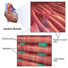
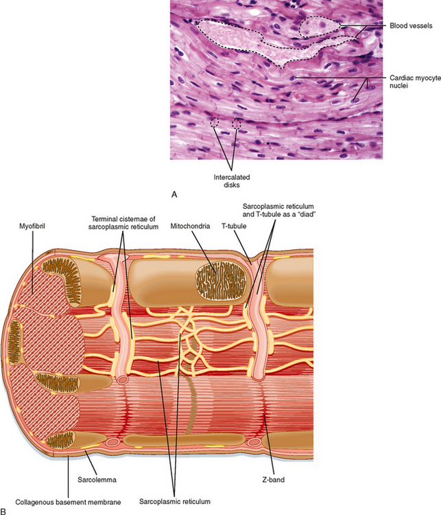
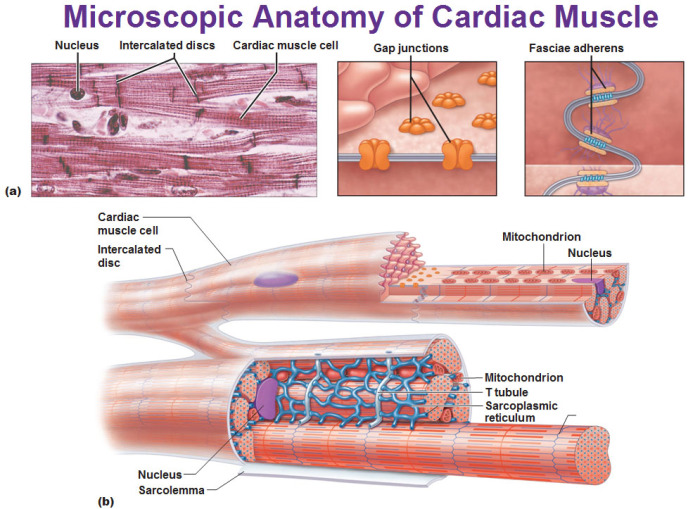
Cardiac muscle tissue diagram. Vor 1 Tag · More information: Soomee Lim et al, Double-layered adhesive microneedle bandage based on biofunctionalized mussel protein for cardiac tissue … Muscle powers the movements of multicellular animals and maintains posture. Its gross appearance is familiar as meat or as the flesh of fish. Muscle is the most plentiful tissue in many animals; for example, it makes up 50 to 60 percent of the body mass in many fishes and 40 to 50 percent in antelopes.Some muscles are under conscious control and are called voluntary muscles. Figure 19.17 Cardiac Muscle (a) Cardiac muscle cells have myofibrils composed of myofilaments arranged in sarcomeres, T tubules to transmit the impulse from the sarcolemma to the interior of the cell, numerous mitochondria for energy, and intercalated discs that are found at the junction of different cardiac muscle cells. (b) A photomicrograph of cardiac muscle cells shows the nuclei and ... 12+ Cardiac Muscle Diagram. Unlike other types of muscle tissue, cardiac myocytes are joined end to end by intercalated. Unlike other muscle cells in the body in the cell, myosin makes up an important group of motor proteins that produce muscular contraction. Broadly considered, human muscle—like the muscles of all vertebrates—is often ...
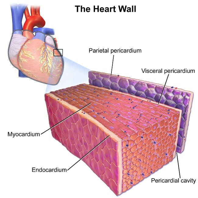
Cardiac muscle tissue, also known as myocardium, is a structurally and functionally unique subtype of muscle tissue located in the heart, that actually has characteristics from both skeletal and muscle tissues.It is capable of strong, continuous, and rhythmic contractions that are automatically generated. The contractility can be altered by the autonomic nervous system and hormones. Muscle tissues are soft tissues that make up the different types of muscles in animals, and give the ability of muscles to contract.It is also referred to as myopropulsive tissue. Muscle tissue is formed during embryonic development, in a process known as myogenesis.Muscle tissue contains special contractile proteins called actin and myosin which contract and relax to cause movement. Cardiac muscle tissue is only found in the heart, and cardiac contractions pump blood throughout the body and maintain blood pressure. Like skeletal muscle, cardiac muscle is striated, but unlike skeletal muscle, cardiac muscle cannot be consciously controlled and is called involuntary muscle. It has one nucleus per cell, is branched, and is distinguished by the presence of intercalated disks. Tissue_types; Muscle; Ultrastructure; Muscle: Skeletal and Cardiac Muscle Ultrastructure. This is a high power, light micrograph of a muscle fibre showing the banding pattern. There are light stripes - which are called the 'Z' lines, and darker wider stripes called the 'A' bands. (A - for anisotropic - because in a polarizing light microscope, the dark bands are birefringent) The Z-lines are ...
Identify the tissue type and its function. Cardiac Muscle •Contracts to propel blood through the circulatory system. Identify the tissue type and its function. Smooth Muscle •Propels substances along internal passageways. Identify the substance that is located inside these holes. Cardiac muscle tissue, or myocardium, is a specialized type of muscle tissue that forms the heart. This muscle tissue, which contracts and releases ...Blood pressure: Systolic mmHgNormal: Less than 120Hypertension stage 2: 140 or higherHypertension stage 1: 130–139 28 Oct 2021 — Cardiac muscle, in vertebrates, one of three major muscle types, ... The heart consists mostly of cardiac muscle cells (or myocardium). A muscle cell is also known as a myocyte when referring to either a cardiac muscle cell (cardiomyocyte), or a smooth muscle cell as these are both small cells. A skeletal muscle cell is long and threadlike with many nuclei and is called a muscle fiber. Muscle cells (including myocytes and muscle fibers) develop from embryonic precursor cells called myoblasts.
The period of timethat begins with contraction of the atria and ends with ventricular relaxation is known as the cardiac cycle (Figure 19.3.1).The period of contraction that the heart undergoes while it pumps blood into circulation is called systole.The period of relaxation that occurs as the chambers fill with blood is called diastole.Both the atria and ventricles undergo systole and diastole ...
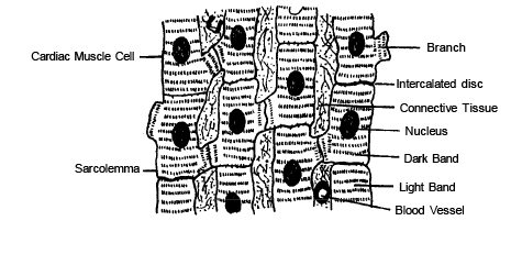
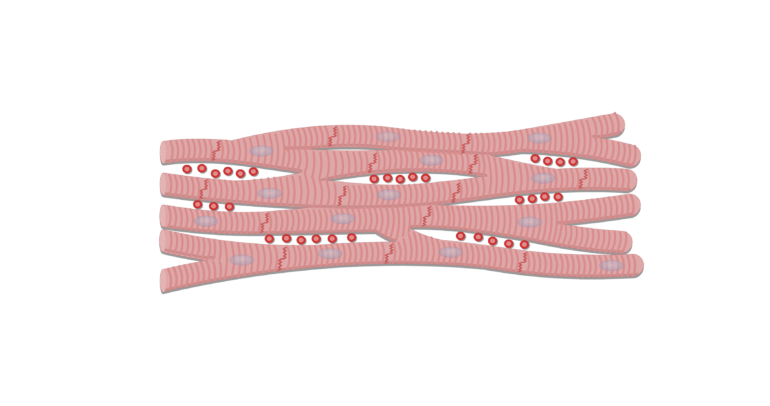
Cardiac muscle fibers cells also are extensively branched and are connected to one another at their ends by intercalated discs. An intercalated disc allows the cardiac muscle cells to contract in a wave-like pattern so that the heart can work as a pump. Cardiac Muscle Tissue. Cardiac muscle tissue is only found in the heart.
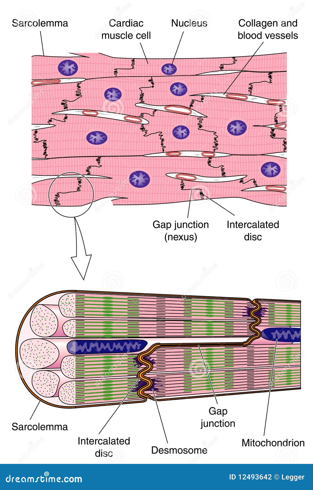
Cardiac Muscle Stock Illustrations 3 663 Cardiac Muscle Stock Illustrations Vectors Clipart Dreamstime
a. allowing blood to flow directly from the right atrium into the left atrium. b. allowing blood to flow directly from the right ventricle into the left ventricle. c. serves as a valvue in the vena cava to regulate venous blood flow. d. prevents back flow of blood from aorta into the left ventricle. e. prevents back flow of blood from pulmonary ...
Write Important Functional Differences Between Striated And Smooth Muscle Tissues Sarthaks Econnect Largest Online Education Community
Structure of Cardiac Muscle. Cardiac muscle is similar to skeletal muscle in that it is striated and that the sarcomere is the contractile unit, with contraction being achieved by the relationship between calcium, troponins and the myofilaments. This article will consider the structure of cardiac muscle as well as relevant clinical conditions.
Cardiac muscle fibers cells also are extensively branched and are connected to one another at their ends by intercalated discs. An intercalated disc allows the cardiac muscle cells to contract in a wave-like pattern so that the heart can work as a pump. Cardiac Muscle Tissue. Cardiac muscle tissue is only found in the heart.
During systole, cardiac muscle tissue is contracting to push blood out of the chamber. Diastole. During diastole, the cardiac muscle cells relax to allow the chamber to fill with blood. Blood pressure increases in the major arteries during ventricular systole and decreases during ventricular diastole.
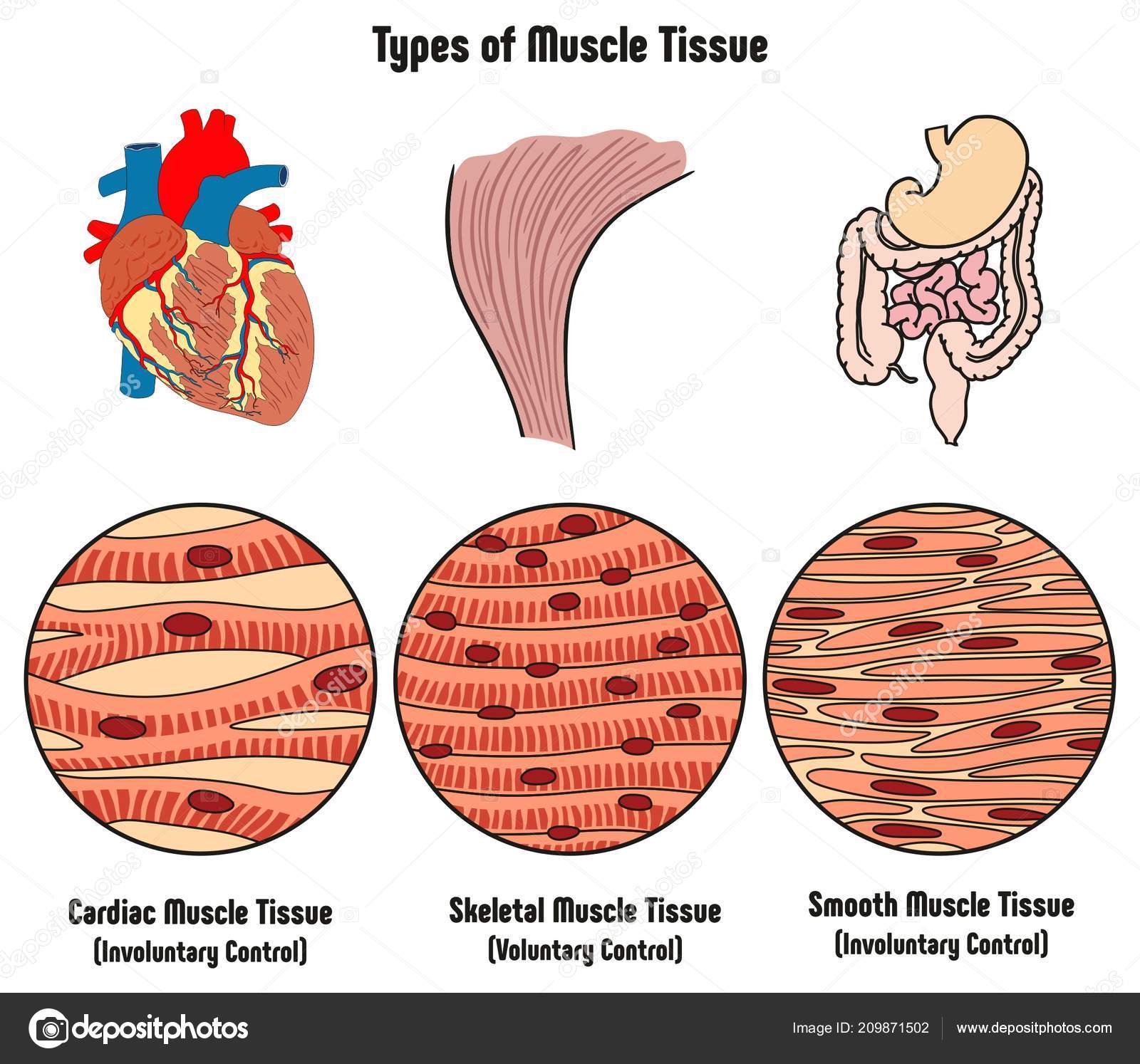
Types Muscle Tissue Human Body Diagram Including Cardiac Skeletal Smooth Stock Vector Image By C Udaix 209871502
28.10.2021 · The cardiac cycle is defined as a sequence of alternating contraction and relaxation of the atria and ventricles in order to pump blood throughout the body. It starts at the beginning of one heartbeat and ends at the beginning of another. The process begins as early as the 4th gestational week when the heart first begins contracting.. Each cardiac cycle has a diastolic phase (also called ...
Cardiac muscle cells usually have a single (central) nucleus. The cells are often branched, and are tightly connected by specialised junctions. The region where ...

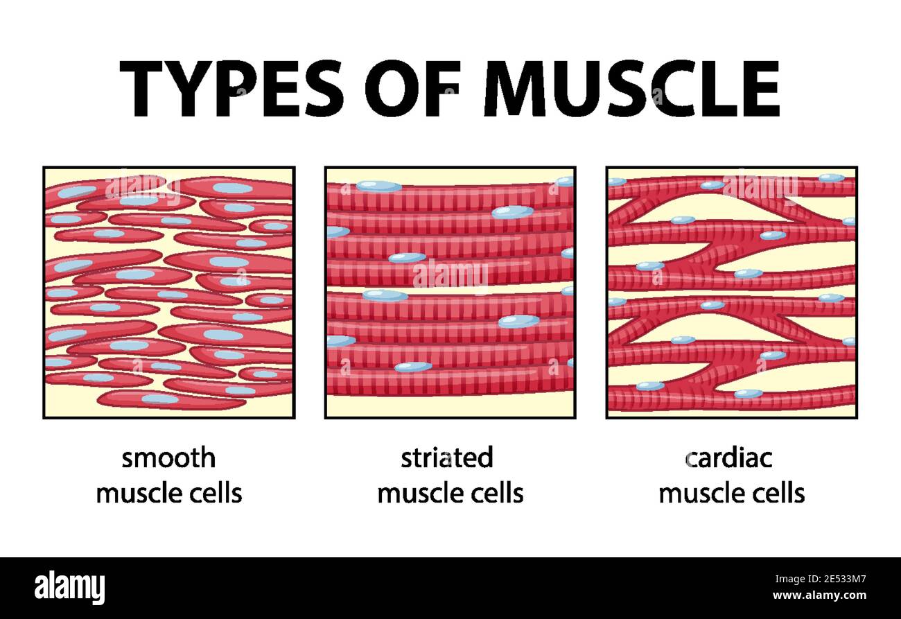
Diagram Showing Types Of Muscle Cells Illustration Royalty Free Cliparts Vectors And Stock Illustration Image 59363058

This Photo Displays Cardiac Muscle Tissue This Tissue Type Is Striated Branched And Has 1 2 N Medical Laboratory Science Tissue Biology Smooth Muscle Tissue

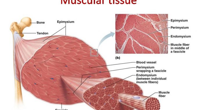






:background_color(FFFFFF):format(jpeg)/images/library/13939/LNOsY5VQ7ADcaM1g9m5g_Cardiac_Muscle.png)

0 Response to "38 cardiac muscle tissue diagram"
Post a Comment