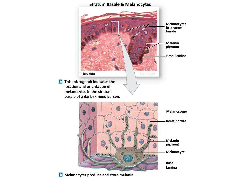
37 drag the labels onto the diagram to identify the melanocyte in the stratum basale of the epidermis.
Start studying Art-labeling Activity: Melanocyte in the Stratum Basale of the Epidermis. Learn vocabulary, terms, and more with flashcards, games, and other study tools. Drag each label to the type of gland it describes. Drag each label to the type of gland it describes.. Label the structures of the hair follicle. Drag each label to the appropriate location on the diagram. Place the following events in the order they occur as a non steroid hormone activates its target cell. Expert answer 100 5 ratings.
Drag the labels onto the diagram to identify the melanocyte in the stratum basale of the epidermis. look at pic The study of tissues using a microscope is called _______________.
Drag the labels onto the diagram to identify the melanocyte in the stratum basale of the epidermis.
Label the components of the integumentary system. Integumentary system parts the skin. For each item below use the pull down menu to select the letter that labels the correct part of the image. Drag the labels onto the diagram to identify the melanocyte in the stratum basale of the epidermis. Large quantities of keratin are found in the ... Drag the labels onto the diagram to identify the melanocyte in the stratum basale of the epidermis. look at pic. Image: Drag the labels onto the diagram to ... Stratum Basale. The stratum basale (also called the stratum germinativum) is the deepest epidermal layer and attaches the epidermis to the basal lamina, below which lie the layers of the dermis. The cells in the stratum basale bond to the dermis via intertwining collagen fibers, referred to as the basement membrane. A finger-like projection, or fold, known as the dermal papilla (plural ...
Drag the labels onto the diagram to identify the melanocyte in the stratum basale of the epidermis.. Module 5.2: The epidermis Epidermal layers overview Entire epidermis lacks blood vessels •Cells get oxygen and nutrients from capillaries in the dermis •Cells with highest metabolic demand are closest to the dermis •Takes about 7-10 days for cells to move from the deepest stratum to the most superficial layer stratum basale d. stratum lucidum g. reticular layer b. stratum corneum e. stratum spinosum h. epidermis as a whole c. stratum granulosum f. papillary layer i. dermis as a whole d; stratum lucidum 1. layer of translucent cells in thick skin containing dead keratinocytes b, d; stratum corneum and lucidum 2. two layers containing dead cells Art-labeling Activities. This activity contains 7 questions. Label the following components in the diagram of the integumentary system. For each item below, use the pull-down menu to select the letter that labels the correct part of the image. 1.1 Epidermis. 1. Stratum Basale or Basal Layer. The deepest layer of the epidermis is called the stratum basale, sometimes called the stratum germinativum. This is where stem cells are located. Because this layer is the innermost layer, many topical products that you apply to the surface of your skin cannot reach this layer and have an effect.
The epidermis is the outermost of the three layers that make up the skin, the inner layers being the dermis and hypodermis. The epidermis layer provides a barrier to infection from environmental pathogens and regulates the amount of water released from the body into the atmosphere through transepidermal water loss.. The epidermis is composed of multiple layers of flattened cells that overlie a ... Question: Part A Drag the labels onto the diagram to identify the layers of the epidermis. Reset stratum basale stratum granulosum stratum lucidum stratum corneum UM straturn spinosum . This problem has been solved! See the answer See the answer See the answer done loading. Show transcribed image text Expert Answer. Who are the experts? Experts are tested by Chegg as specialists in their ... 1. Stratum basale (basal layer).The stratum basale consists of a single layer of cells in contact with the dermis.Four types of cells compose the stratum basale: keratinocytes, melanocytes, tactile cells (Merkel cells), and nonpigmented granular dendrocytes (Langerhans cells).With the exception of tactile cells, these cells are constantly dividing mitotically and moving outward to renew the ... Transcribed image text: Modules MasteringAandP Mastering Course Home (Click here for HOMEWORK, and TESTS) Ch 05 HW Art-labeling Activity: Melanocyte in the Stratum Basale of the Epidermis 5 of 15 rart A Drag the labels onto the diagram to identity the melanocyle in the stratum basale of the epidermis Reset Helo Statum basae Demis Melann pgment Basement mentrane Keratinocye Melanosome Melaocyle ...
23.5.2020 · 109 Likes, 2 Comments - Dr Raymond C Lee MD (@drrayleemd) on Instagram: “What an amazing virtual aats. Congratulations to my chairman Dr Vaughn Starnes 100th AATS…” The layers of the epidermis include the stratum basale (the deepest portion of the epidermis), stratum spinosum, stratum granulosum, stratum lucidum, and stratum corneum (the most superficial portion of the epidermis). Stratum basale, also known as stratum germinativum, is the deepest layer, separated from the dermis by the basement membrane (basal lamina) and attached to the basement membrane ... Figure 5.2 The main structural features of the skin epidermis. MelanocyteDesmosomes Melanin granule Tactile (Merkel) cell Sensory nerve ending Epidermal dendritic cell Dermis Dermis Keratinocytes (b) (a) Stratum corneum Stratum granulosum Stratum spinosum Stratum basale Part A Drag the labels onto the diagram to identify the main structural features in the epidermis of thin skin. Reset Help Melanocyte Melanocyte Stratum corneum ...
Question: Part A Drag the labels onto the diagram to identify the melanocyte in the stratum basale of the epidermis. Reset Help Melanin pigment Basement ...

The Stoddart Visual Dictionary Look Up The Word From The Picture Find The Picture From The Word Stoddart Publishing Co Ltd Jean Claude Corbeil Pdf Fruit Moon
55. Sudoriferous (sweat) glands are categorized as two distinct types. Which of the following are the two types of sweat glands? eccrine and apocrine. 56. Which layer (s) of the skin is (are) damaged in a second-degree burn? The epidermis and the superficial region of the dermis are damaged. 57.
Drag the labels onto the diagram to identify the cells and fibers of connective tissue proper using diagrammatic and histological views. ... Drag the labels onto the diagram to identify the melanocyte in the stratum basale of the epidermis. ... the entire epidermis and perhaps some of …
Anatomy of the Skin. The skin is a vital organ that covers the entire outside of the body, forming a protective barrier against pathogens and injuries from the environment. The skin is the body's largest organ; covering the entire outside of the body, it is about 2 mm thick and weighs approximately six pounds.
Drag each label into the appropriate position to identify which chemical classification it describes. In meat we know it as gristle. Label the microscopic features of the pancreas. Drag each label to the cell type it describes. Place the following events in the order they occur as a non steroid hormone activates its target cell.
Academia.edu is a platform for academics to share research papers.
Anatomy 3 2 Integumentary System Epidermis Labeling Diagram Quizlet Play this quiz called label the integumentary system and show off your skills. Label integumentary system. Start studying integumentary. Question: A. Identify And Label The Skin Model Structures Numbers 1 To 8. This problem has been solved! See the answer. Show transcribed ...
Stratum basale, and stratum spinosum. The basal cell layer (stratum basale, or stratum germinosum), is a single layer of cells, closest to the dermis. It is usually only in this layer that cells divide. Some of the dividing cells move up to the next layer. The prickle cell layer (stratum spinosum) is the next layer (8-10 layers of cells). The ...
Skin tissue engineering. David J. Wong 1, Howard Y. Chang 1, §. 1 Program in Epithelial Biology, Stanford University, Stanford, CA 94305. The skin is the largest organ of the body and is critical to survival of the organism as a barrier to the environment and for thermal regulation and hydration retention.
Drag the labels onto the diagram to identify the melanocyte in the stratum basale of the epidermis. Basement Membrane. Melansome. Keraitnocyte.
Solved Utural Stoll Inc Skin Question 11 Yes X No Analyse The Image Below And Label The Layers And Cells Of The Skin C K D H M G K Holoski F Course Hero
A. Tactile cells anchor the skin to the body. B. Keratinocytes produce a fibrous protein to protect the epidermis. C. Melanin provides protection against ultraviolet (UV) radiation. D. Langerhans cells activate the immune system. A. Tactile cells anchor the skin to the body. The skin consists of two main regions.
Epidermis. The epidermis is composed of keratinized, stratified squamous epithelium. It is made of four or five layers of epithelial cells, depending on its location in the body. It is avascular.. Thin skin has four layers of cells. From deep to superficial, these layers are the stratum basale, stratum spinosum, stratum granulosum, and stratum corneum.Most of the skin can be classified as thin ...
Identify the epidermis, dermis and determine the type of tissue in each ... and label the dermal papilla and dermis •Name the Layers of skin and label the ... • SC: Stratum corneum SS: Stratum spinosum • SL: Stratum lucidum SB: Stratum basale • SG: Stratum granulosum SG. Identify the thick and thin skin. Can you name the layers ...
Generally, in the big schema things of the human body, the skin often does not strike as an organ. However, the skin is composed of tissues and performs mission-critical functions in the body.. The skin and their accessory structures such as hair, glands, and nails make up the integumentary system, which provides the body with overall protection.. The skin contains multiple layers of cells and ...
Transcribed image text: - Parta Drag the labels onto the diagram to identity the melanocyte in the stratum basale of the epidermis. Reset Help Demon IN We.
Part A Drag the labels onto the diagram to identify the components of the integumentary system. ANSWER: Help Reset Stratum basale Dermis Basement membrane Melanocyte Melanin pigment Keratinocyte Melanosome. Correct Art-labeling Activity: Components of the Integumentary System, Part 2 Learning Goal: To learn the components of the integumentary system. Label the components of the integumentary ...
Stratum Basale. The stratum basale (also called the stratum germinativum) is the deepest epidermal layer and attaches the epidermis to the basal lamina, below which lie the layers of the dermis. The cells in the stratum basale bond to the dermis via intertwining collagen fibers, referred to as the basement membrane. A finger-like projection, or fold, known as the dermal papilla (plural ...
In most regions of the body the epidermis has four strata or layers —stratum basale, stratum spinosum, stratum granulosum, and a thin stratum corneum. This is called thin skin. Where exposure to friction is greatest, such as in the fingertips, palms, and soles, the epidermis has five layers—stratum basale, stratum spinosum, stratum ...
Keratinocytes of the stratum corneum start producing a thick extracellular layer of keratin fibers, which surround the cell, making it impermeable to nutrients. The cell eventually dies and becomes the dead keratinized cell of the stratum corneum. After the stem cells of the basale layer divide, the daughter cells migrate upwards to the stratum ...
Dr Vikram Sood Clinic Hair Transplant In Jalandhar Fue In Punjab Hair Fall Treatment In Jalandhar Hair Transplant In Punjab
Drag the labels onto the diagram to identify the main structural features in the epidermis of thin skin. Who are the experts? Experts are tested by Chegg as specialists in their subject area. We review their content and use your feedback to keep the quality high. Answer : IF ….
Stratum Basale. The stratum basale (also called the stratum germinativum) is the deepest epidermal layer and attaches the epidermis to the basal lamina, below which lie the layers of the dermis. The cells in the stratum basale bond to the dermis via intertwining collagen fibers, referred to as the basement membrane. A finger-like projection, or fold, known as the dermal papilla (plural ...
Drag the labels onto the diagram to identify the melanocyte in the stratum basale of the epidermis. look at pic. Image: Drag the labels onto the diagram to ...
Label the components of the integumentary system. Integumentary system parts the skin. For each item below use the pull down menu to select the letter that labels the correct part of the image. Drag the labels onto the diagram to identify the melanocyte in the stratum basale of the epidermis. Large quantities of keratin are found in the ...


0 Response to "37 drag the labels onto the diagram to identify the melanocyte in the stratum basale of the epidermis."
Post a Comment