40 figure 11-4 is a diagram of the frontal section of the heart
PDF PowerPoint Lecture Slides prepared by Barbara Heard ... Figure 18.9 The heart is a double pump, each side supplying its own circuit. Both sides of the heart pump at the same time, but let's follow one spurt of blood all the way through the system. Oxygen-rich blood Superior vena cava (SVC) Inferior vena cava (IVC) Coronary sinus Right atrium Tricuspid valve Pulmonary Semilunar Right valve ... Figure 35.3 Frontal Section of Human Heart Diagram | Quizlet Start studying Figure 35.3 Frontal Section of Human Heart. Learn vocabulary, terms, and more with flashcards, games, and other study tools.
PDF BIO 113 LAB 1. Anatomical Terminology, Positions, Planes ... BIO 113 Fall 2011 LAB 1 Page 11 Visualizing a section in the 90 degree plane: Reconstruction The sections you have just drawn are in a plane known as longitudinal. You will now use these sections to create a cross section. For this assignment you should choose a plane crossing each of your sections in which there is some of the egg yolk present.

Figure 11-4 is a diagram of the frontal section of the heart
Heart Anatomy Frontal Section Diagram | Quizlet The first major branch off of the aorta and the major artery to the forelimbs and head. Superior Vena Cava. receives blood from the head and arms and chest and empties into the right atrium of the heart. Ascending Aorta. the ascending part of the aorta as it emerges from the left ventricle. Pulmonary Semilunar Valve. 33 Label This Anterior View Of The Human Heart - Labels ... Figure 4410 identify the features indicated on this anterior view of a frontal section of a human heart model using the terms provided. Correctly label the following anatomical features of the heart and thoracic cage correctly label the following vessels leading from and toward the anterior heart correctly label the following external anatomy ... A Labeled Diagram of the Human Heart You Really Need to ... A Labeled Diagram of the Human Heart You Really Need to See. The heart, one of the most significant organs in the human body, is nothing but a muscular pump which pumps blood throughout the body. The human heart and its functions are truly fascinating. The heart, though small in size, performs highly significant functions that sustains human life.
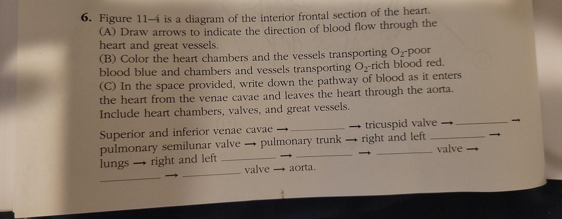
Figure 11-4 is a diagram of the frontal section of the heart. DOC Dorchester School District Two / Homepage Figure 11-4 is a diagram of the frontal section of the heart. Follow the instructions below to complete this exercise. (a) Correctly identify each of the heart valves (numbers 6-9 on the figure) by inserting the appropriate terms in the blanks left of the figure. (b) Use the numbers from the figure to identify the structures described below. PDF ANATOMICAL TERMINOLOGY: Body Planes and Sections 4. _____- vertical cut at right angles to sagittal plane, divides the body into anterior and posterior portions 5. _____- cross-section, a horizontal cut that divides the body into upper and lower parts Identify the planes or sections on the diagram. Use the terms to indicate the plane or section. 6. Cardiovascular Coloring Packet 2019.doc - Ch 11 ... Figure 11-4 is a diagram of the frontal section of the heart. Follow the instructions below to complete this exercise. (a) Correctly identify each of the heart valves (numbers 6-9 on the figure) by inserting the appropriate terms in the blanks left of the figure. (b) Use the numbers from the figure to identify the structures described below. Solved Chapter 11 The Cardiovascular System 181 6. Figure ... Figure 11-4 is a diagram of the frontal section of the heart. Follow the instructions below to complete this exercise. First, draw arrows to indicate the direction of blood flow through the heart. Draw the pathway of the oxygen-rich blood with red arrows, and trace the pathway of oxygen-poor blood with blue arrows. This problem has been solved!
PDF Castle High School - A Warrick County High School Figure 7—7 shows a frontal view of the meninges of the brain at the level of the superior sagittal (dural) sinus_ First, labet the arachnoid Villi on the figure. and diff,- to co}or the coding circles and corresponding structures in the diagram. Dura mater Arachnoid mater Scalp Bone of skuiÍ Superior sagittal sinus Gray matter of cerebral cortex Diagram of Human Heart and Blood Circulation in It | New ... Four Chambers of the Heart and Blood Circulation. The shape of the human heart is like an upside-down pear, weighing between 7-15 ounces, and is little larger than the size of the fist. It is located between the lungs, in the middle of the chest, behind and slightly to the left of the breast bone. The heart, one of the most significant organs ... PDF The Cardiovascular System - Pearson of the blood vessels leaving and entering the heart. (Figure 11.3 shows two views of the heart—an exter-nal anterior view and a frontal section. As the ana-tomical areas of the heart are described in the next section, keep referring to Figure 11.3 to locate each of the heart structures or regions.) Chambers and Associated Great Vessels 7.3 The Skull - Anatomy & Physiology Important landmarks of the temporal bone, as shown in Figure 7.3.7, include the following:. External acoustic meatus (ear canal)—This is the large opening on the lateral side of the skull that is associated with the ear.; Internal acoustic meatus—This opening is located inside the cranial cavity, on the medial side of the petrous ridge.It connects to the middle and inner ear cavities of ...
Blank Diagram Of Heart To Label Figure 11-4 is a diagram of the frontal section of the point Follow the. There are for printing is a label blank diagram of heart to eat. Heart diagram blank as most popular color You guessed it... Cancellation of Electrocardiographic Effects During ... FIGURE 1 Diagrammatic representation of the construction of a frontal plane section (B) from horizontal plane cardiac sections (A). The points labeled 1 through 8 on the horizontal sections illustrate the measurements carried out to construct frontal plane sections. These points are shown on the frontal section in their appropriate relation to ... Brain | Biology for Majors II - Lumen Learning There are three different ways that a brain can be sectioned in order to view internal structures: a sagittal section cuts the brain left to right, as shown in Figure 1b, a coronal section cuts the brain front to back, as shown in Figure 1a, and a horizontal section cuts the brain top to bottom. Cerebral Cortex PDF Chapter 18 The Heart - Mrs. Ahrens' Science Site The figure below is a diagram of the frontal section of the heart. Follow the instructions below to complete this exercise, which considers both anatomical and physiological aspects of the heart. 1) Draw arrows to indicate the direction of blood flow through the heart. Draw the pathway of the oxygen-rich
3 Sketch of superior frontal section showing scalp, skull ...
PDF The Cardiovascular 11 CHAPTER OUTLINE System The adult heart is shown in Figure 11.4 Note the thick ventricular walls, especially in the left ventricle. It is the left ventricle that must generate enough force to push blood throughout the body. The less muscular right ventricle pushes blood only to the nearby lungs.

Comparative analysis of the combined petrosal and the ...
19.1 Heart Anatomy - Anatomy & Physiology A transverse section through the heart slightly above the level of the atrioventricular septum reveals all four heart valves along the same plane (Figure 19.1.11). The valves ensure unidirectional blood flow through the heart. Between the right atrium and the right ventricle is the right atrioventricular valve, or tricuspid valve. It typically ...

SARS‐CoV2 disease seen through the prism of acutely ...
Solved 6. Figure 11-4 is a diagram of the interior frontal ... Figure 11-4 is a diagram of the interior frontal section of the heart. (A) Draw arrows to indicate the direction of blood flow through the heart and great vessels. (B) Color the heart chambers and the vessels transporting O2-poor blood blue and chambers and vessels transporting O2-rich blood red.
Thesis Reference
PDF M11 MARI5326 12 SE C11.indd Page 356 24/09/16 9 ... - Pearson of the blood vessels leaving and entering the heart. (Figure 11.3 shows two views of the heart—an exter-nal anterior view and a frontal section. As the ana-tomical areas of the heart are described in the next section, keep referring to Figure 11.3 to locate each of the heart structures or regions.) Chambers and Associated Great Vessels

19.1 Heart Anatomy – Anatomy & Physiology
PDF Home - Buckeye Valley Created Date: 1/23/2014 12:31:42 PM

Impact of Cardiac Resynchronization Therapy on Heart ...
CARDIOVASCULAR WORKBOOK OLA.docx - The Cardiovascular ... Figure 11—4 is a diagram of the frontal section of the heart. Follow the instructions below to complete this exercise. The colored arrows indicate the flow of oxygenated (red) and deoxygenated (blue) blood through the heart.

14 Heart disease ideas | chd awareness, heart disease ...
PDF Anatomy of the Heart Anatomy of the Heart Objectives: 1. To describe the location of the heart. 2. To name and locate the major anatomical areas and structures of the heart when provided with an appropriate model or diagram, or with a dissected sheep heart, and to explain the function of each. 3. To trace the pathway of blood through the heart. 4.
Anatomy & Physiology Coloring A Complete Study Guide Twelfth ...
Ch. 14 Introduction - Anatomy and Physiology - OpenStax Ch. 14 Introduction - Anatomy and Physiology | OpenStax. Figure 14.1 Too Hot to Touch When high temperature is sensed in the skin, a reflexive withdrawal is initiated by the muscles of the arm. Sensory neurons are activated by a stimulus, which is sent to the central nervous system, and a motor response is sent out to the skeletal muscles that ...

3 Figure 11 2 is an anterior view of the heart Identify each ...
Anatomy of the normal fetal heart: The basis for ... (c) Sagittal section of a 28-week fetal heart showing the coronary sinus which extends through the left atrioventricular groove and is separated from the walls of the LA. (d-f) Frontal sections of a 23-week heart, where image e is a histologic stain using trichromic Masson stain corresponding to image d.

Adventitial elastolysis is a primary event in aneurysm ...
PDF Brain Review and Wkst Answer Keys - Mayfield City School ... Created Date: 4/30/2013 4:05:46 PM
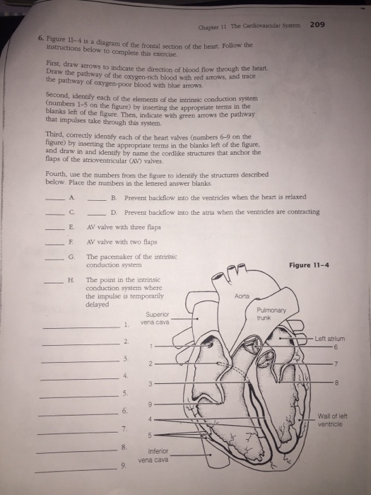
Solved 209 Chapter 11 The Cardiovascular System 6. Figure ...
PDF Brain Anatomy - Wou BI 335 - Advanced Human Anatomy and Physiology Western Oregon University Figure 4: Mid-sagittal section of brain showing diencephalon (includes corpus callosum, fornix, and anterior commissure) Marieb & Hoehn (Human Anatomy and Physiology, 9th ed.) - Figure 12.10 Exercise 2: Utilize the model of the human brain to locate the following structures / landmarks for the
Genetics of Skin Cancer (PDQ®)–Health Professional Version ...
A Labeled Diagram of the Human Heart You Really Need to ... A Labeled Diagram of the Human Heart You Really Need to See. The heart, one of the most significant organs in the human body, is nothing but a muscular pump which pumps blood throughout the body. The human heart and its functions are truly fascinating. The heart, though small in size, performs highly significant functions that sustains human life.
Untitled
33 Label This Anterior View Of The Human Heart - Labels ... Figure 4410 identify the features indicated on this anterior view of a frontal section of a human heart model using the terms provided. Correctly label the following anatomical features of the heart and thoracic cage correctly label the following vessels leading from and toward the anterior heart correctly label the following external anatomy ...

Activation of Peripheral and Central Trigeminovascular ...
Heart Anatomy Frontal Section Diagram | Quizlet The first major branch off of the aorta and the major artery to the forelimbs and head. Superior Vena Cava. receives blood from the head and arms and chest and empties into the right atrium of the heart. Ascending Aorta. the ascending part of the aorta as it emerges from the left ventricle. Pulmonary Semilunar Valve.

Cardiovascular system - ppt download
Solved 6. Figure 11-4 is a diagram of the interior frontal ...
Name: Period:
Integrative genetic analysis of the amyotrophic lateral ...
931914 - Repetitive Transcranial Magnetic Stimulation (rTMS ...

Frontal Section of the Heart Diagram Diagram | Quizlet
Cells | Free Full-Text | Interaction of Mitochondrial Calcium ...

Light-induced asymmetries in the embryonic retina are ...

Lab Fig 3.4 Frontal section of the heart Diagram | Quizlet
Rare variants in the endocytic pathway are associated with ...
Heart Rhythm By Hiren Kevadiya

Neural correlates of audio-visual integration of socially ...

53 Atrial Septal Defect (ASD) ideas | atrial septal defect ...

The anatomy of the human frontal lobe - ScienceDirect
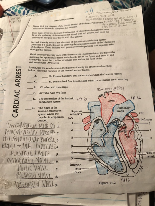
Solved ani &10 Dr nnana tdiagram of the frontal section of ...
Untitled
Fiber Pathways of the Brain | Semantic Scholar
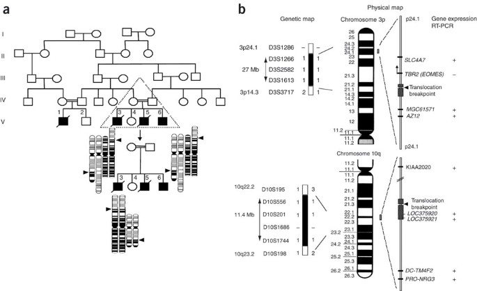
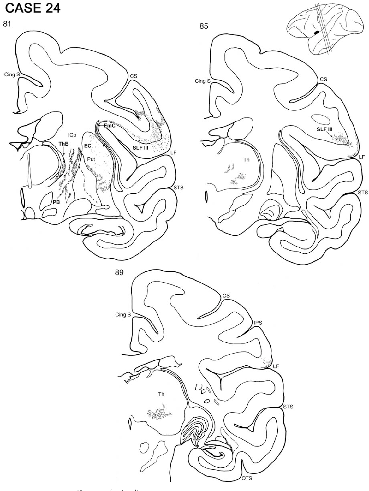
Homozygous silencing of T-box transcription factor EOMES ...
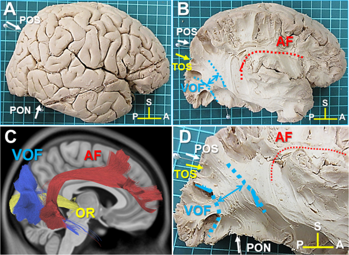
White matter dissection and structural connectivity of the ...

Sex differences in vectorcardiogram of African-Americans with ...
Brain Sciences | Free Full-Text | Bi-Temporal Anodal ...

Acupuncture Decreases Blood Pressure Related to Hypothalamus ...

UGA Anatomy and Physiology 2 Lab Manual

The anatomy of the human frontal lobe - ScienceDirect

Frontal Section through the heart Diagram | Quizlet
Untitled
0 Response to "40 figure 11-4 is a diagram of the frontal section of the heart"
Post a Comment